Carbohydrates for the MCAT: Everything You Need to Know
/Master MCAT carbohydrate concepts including monosaccharides, disaccharides, polysaccharides, glycolysis, and glycogen metabolism. Includes practice questions with answers.
(Note: This guide is part of our MCAT Biochemistry series.)
Part 1: Introduction to RNA
Part 2: Structure of RNA
a) Similarities to DNA
b) Differences with DNA
Part 3: Functions of RNA
a) mRNA
b) tRNA
c) rRNA
d) snRNA
Part 4: High-yield terms
Part 5: Passage-based questions and answers
Part 6: Standalone questions and answers
----
Part 1: Introduction
Although modern humans have been around for at least 100,000 years, one topic that seems to puzzle us to this day is our diet. In grocery stores all across the United States, you’ll find plenty of food products marketed as “low-carb” or “keto-friendly.” You may have even heard of famous athletes, such as Lebron James and Tim Tebow, sharing their results on a low-carb diet.
Despite the bad press, carbohydrates certainly played—and continue to play—a significant role in our evolutionary history and shaped who we are today. Thus, it’s no surprise that you are expected to understand them in detail as you pursue a career in medicine. In this article, we’ll discuss the basic biochemistry behind carbohydrates so that you can have a strong foundation to build on when reviewing their metabolism.
Without further ado, let’s begin!
----
Part 2: Useful reactions
a) Fischer and Haworth projections
Carbohydrates, also commonly referred to as “sugars,” can be represented in many forms. One such form is a Fischer projection, a two-dimensional representation of a molecule that gives three-dimensional information. The horizontal lines of a Fischer projection are equivalent to wedges: functional groups on horizontal lines are coming out of the page. The vertical lines are equivalent to dashes: functional groups on vertical lines are going into the page.
Figure: Fischer projections of D- and L- forms of glyceraldehyde.
For sugars, the absolute configuration is designated using D- and L- nomenclature instead of the R and S system used in organic chemistry. If the hydroxyl group of the highest-numbered chiral carbon is on the right, the sugar is in the D-configuration. If the hydroxyl group of the highest-numbered chiral carbon is on the left, it is in the L-configuration.
While Fischer projections represent the straight-chain form of carbohydrates, you may also see sugars represented in their cyclic form as Haworth projections. In a cyclic sugar, the anomeric carbon is the carbon that has two bonds to oxygen.
Be aware that a cyclic sugar can exist in two possible anomers: an ⍺-anomer and a β-anomer. A sugar is in its α-anomer form when the anomeric carbon’s substituent is on the opposite side of the plane as the highest numbered chiral center’s substituent. A sugar is in its β-anomer form when the anomeric carbon’s substituent is on the same side of the plane as the highest numbered chiral center’s substituent.
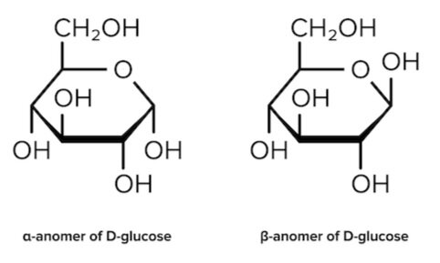
Figure: Haworth projections of the α- and β-anomers of D-glucose.
b) Cyclization
Cyclic sugars are formed via an intramolecular reaction, or a reaction between two functional groups on the same molecule. Aldoses are sugars with an aldehyde group (-CHO). Their cyclization forms a hemiacetal. On the other hand, Ketoses have a ketone group (-CO) and no other further oxidized functional groups. Their cyclization forms a hemiketal.
It’s easiest to visualize this reaction when the carbohydrate is drawn in its linear form (e.g., in a Fischer projection). During cyclization of both aldoses and ketoses, the hydroxyl group (nucleophile) on the highest-numbered chiral center attacks the carbonyl group (electrophile).
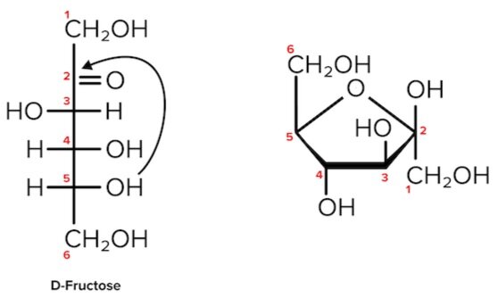
Figure: Cyclization of D-fructose, a hemiketal. The second carbon is both the site of nucleophilic attack and the anomeric carbon (as it is bonded to 2 oxygen atoms).
Note that as we convert the structure of the sugar from its two-dimensional to three-dimensional structure, the functional group that is pointing to the right of the Fischer projection will end up pointing downward in the Haworth projection.
For most sugars that are dissolved in a solution, this cyclization occurs spontaneously. As a result, many sugars can be found in various combinations of D-, L-, and linear forms. (The final ratios of each of these forms are usually affected by stereochemistry unique to each molecule.) This tendency to undergo spontaneous cyclization is known as mutarotation.
Sugars can also be described as being “non-reducing” or “reducing.” A reducing sugar is one that can act as a reducing agent. Reducing sugars can be identified through the presence of a free anomeric carbon, meaning it is not in a glycosidic bond and has a free hydroxyl group. Conversely, non-reducing sugars lack a free anomeric carbon.
Benedict’s reagent and Tollen’s reagent are two reagents that are commonly used to detect the presence of reducing sugars. The two reagents react with reducing functional groups in unique ways: Benedict’s reagent reacts with aldoses to form a red copper precipitate, while Tollen’s reagent reacts with aldehydes to form a silver, mirrorlike precipitate.
----
Part 3: Sugar structures
A sugar monomer is referred to as a monosaccharide. Two monosaccharides can be attached via a glycosidic linkage to form a disaccharide. Multiply monomers that are linked together form a polysaccharide.
All monosaccharides are considered reducing sugars. Sucrose, a disaccharide we will discuss below, is a common example of a non-reducing sugar.
All monosaccharides, regardless of form, share a common chemical composition:
One part carbon atoms
Two parts hydrogen atoms
One part oxygen atoms
This composition varies slightly in disaccharides and polysaccharides but holds true for the composition of all monosaccharides.
The MCAT will test your knowledge of how these sugars behave in reactions and also on the structure of particular monosaccharides and disaccharides. Some of these structures are discussed below.
a) High-yield monosaccharides
One of the most common monosaccharides you will encounter is glucose. This is an aldose sugar, and in its cyclic form, it is classified as a pyranose, meaning it is a six-membered ring.
Another common sugar you may see is fructose. This monosaccharide is a ketose sugar, and in its cyclic form, it is classified as a furanose, meaning it is a five-membered ring. (You may also see certain sugars referred to as hexoses and pentoses, meaning they have six and five carbon atoms, respectively. Note that it’s possible for a sugar to be both a hexose and a furanose—the sugar would simply have five atoms within the ring and at least one carbon atom outside of the ring.)
Galactose, like glucose, is another aldose sugar. However, this sugar has four cyclic isomers, with two being pyranoses and the other two being furanoses.
Gain instant access to the most digestible and comprehensive MCAT content resources available. 60+ guides covering every content area. Subscribe today to lock in the current investments, which will be increasing in the future for new subscribers.
Figure: Study these monosaccharides well! You should expect to be tested on them.
Oxyribose, or ribose, is a monosaccharide that is a constituent of RNA. Deoxyribose, a relative of ribose, is a constituent of DNA. A deoxy sugar is a ribose that has replaced one hydroxyl group with a hydrogen atom. In RNA, the 2’ carbon has a hydroxyl substituent, whereas in DNA, the 2’ carbon has this replaced with a hydrogen atom.
Figure: Ribose and deoxyribose differ by one hydroxyl (-OH) group.
You should be able to recognize and differentiate the structures of the pentose sugars found in DNA and RNA.
b) High-yield disaccharides
In addition to the above monosaccharides, it will be useful to memorize the structures of certain disaccharides. Here’s a helpful hint: these disaccharides will be formed by combinations of the monosaccharides you already know! Recall that monosaccharides can be attached to each other through glycosidic bonds.
Sucrose is formed via the α-1,2 glycosidic linkage of an α-glucose molecule and a β-fructose molecule. This means that an α-bond is formed between the 1st and 2nd carbons of glucose and fructose, respectively.
Lactose is formed via the β-1,4 glycosidic linkage of a β-galactose and β-glucose
Maltose is formed via the α-1,4 glycosidic linkage of two α-glucose molecules.
Figure: The structures of high-yield disaccharides.
c) Glycogen
While monosaccharides and disaccharides are the most usable form of energy, they can be highly inefficient to store. As a result, our bodies store carbohydrates in the form of a polysaccharide known as glycogen.
Glycogen is a polysaccharide that is highly branched and compacted—to maximize storage capacity and minimize the solvation energy needed to store this molecule.
Starting with a peptide core, an enzyme known as glycogen synthase linearly arranges the glucose molecules via α-1,4 glycosidic links. Another enzyme known as branching enzyme hydrolyzes one of these linkages and starts a new branch using the oligosaccharide and attaching it via an α-1,6 glycosidic linkage. Multiple glycogen chains are arranged in a spherical fashion to form a granule. This process of glycogen formation is known as glycogenesis.
In the reverse process, known as glycogenolysis, the large glycogen molecule is broken down into its smaller glucose units. An enzyme known as glycogen phosphorylase breaks the α-1,4 glycosidic linkages in a linear chain until it reaches the branching point. Here, the debranching enzyme will hydrolyze the α-1,4 glycosidic linkage and relocate the oligosaccharide to the end of another linear chain. It will then hydrolyze an α-1,6 glycosidic linkage between the branched glucose molecule and the linear chain.
Note that two enzymes are required for both glycogenesis and glycogenolysis: one to create/break down glucose molecules in a linear chain, and another to branch/debranch a chain of existing glucose molecules.
For further information on the fate of glucose and its pathways, refer to our guide on carbohydrate metabolism.
----
Part 4: High-yield terms
Fischer projection: a two-dimensional representation of a molecule that gives three-dimensional information; represents sugars in D- and L-configurations
Haworth projection: a representation of the cyclic forms of sugars; represents sugar in their ⍺-anomer or β-anomer form
Aldose: a sugar with an aldehyde group (-CHO); cyclization forms a hemiacetal
Ketose: a sugar with a ketone group (-CO); cyclization forms a hemiketal
Mutarotation: the ability of monosaccharides in aqueous solution to undergo spontaneous cyclization
Pyranose: a sugar that contains a six-membered ring; e.g., glucose
Furanose: a sugar that contains a five-membered ring; e.g., fructose
Benedict’s reagent: reacts in the presence of aldoses to form a red copper precipitate
Tollen’s reagent: reacts in the presence of aldehydes to form a silver, mirrorlike precipitate
Glycogen: a polysaccharide used by the body as its primary form of carbohydrate-based energy storage
----
Part 5: Passage-based questions and answers
Congenital defects in metabolic pathways can result in chronic deficiencies that persist through infancy and adulthood. A congenital deficiency of the metabolic enzyme α-glucosidase (GAA) leads to infantile-onset Pompe disease (IOPD). This disease results in the buildup of lysosomal glycogen in tissues such as the skeletal muscle and heart, along with risk of heart failure in early infancy.
Glycogen is typically located in the cell cytoplasm, with only a small amount of glycogen present in the lysosomes. In one experiment, researchers collected and analyzed myocytes derived from the pluripotent stem cells of three IOPD patients (Pom1, Pom2, Pom3) to investigate the pathophysiology of the disease and possible therapeutic interventions. A glucose-free culture medium was used to establish cell cultures. A periodic acid-Schiff stain was conducted to compare the amounts of glycogen in IOPD-derived myocytes and control. The results are shown in Figure 1.
Figure 1: Ctr - control; Pom - experimental condition
Enzyme replacement therapy with recombinant human GAA (rhGAA) has been shown to improve the survival rate in IOPD patients. In a second experiment, researchers added rhGAA at three different concentrations to 3 previously established cell cultures and recorded the amount of glycogen present in cells. The results are shown in Figure 2.
Question 1: Glucosidases break down complex carbohydrates, such as glycogen. Which of the following enzymes is a glucosidase?
A) Glycogen synthase
B) Phosphofructokinase I
C) Debranching enzyme
D) Branching enzyme
Question 2: Why did the researchers use a glucose-free culture medium in experiment 1?
A) To promote the production of ketones
B) To ensure that only lysosomal glycogen would be detected
C) To improve the accuracy of periodic acid-Schiff stains
D) None of the above
Question 3: Scientists discover that when rhGAA at a concentration of 35 nM is added to the Pom3 cell line, the amount of glycogen detected is equal to 40% less the baseline value. Which of the following conclusions can be drawn?
A) The experiment has been invalidated due to contamination and must be performed again
B) The effects of rhGAA are dose-dependent
C) The measurements taken from the Pom2 line are statistically significant
D) The effects of rhGAA are dose-independent
Question 4: Which of the following accurately describes the types of cells the scientists used to generate the myocytes for their research?
A) They can differentiate into any fetal or placental cell type
B) They can only differentiate into any cell type found in the placental structures
C) They can only differentiate into the cell types of one tissue or organ
D) They can differentiate into any of the cell types found in the three embryonic germ layers but not into those found in the placental structures
Question 5: In humans, glycogen is stored in the form of:
A) Triglycerides
B) Chylomicrons
C) Starch
D) Granules
Answer key for passage-based questions
Answer choice C is correct. Debranching enzyme is essential in the breakdown of glycogen (choice C is correct). Glycogen synthase catalyzes the polymerization of glucose monomers into linear chains of glycogen (choice A is incorrect). Phosphofructokinase I catalyzes the addition of a phosphate to fructose 6-phosphate during glycolysis (choice B is incorrect). Branching enzyme is a key enzyme in glycogenesis that creates the branches found in glycogen (choice D is incorrect).
Answer choice B is correct. The passage states that glycogen is primarily found in the cytoplasm and only a small amount is in the lysosome. Since the experiment seeks to quantify lysosomal glycogen accumulation, the researchers must ensure that they are not staining cytoplasmic glycogen. Deprivation of glucose would lead to the consumption of cytoplasmic glycogen and allow researchers to only stain lysosomal glycogen (choice B is correct). The researchers are not interested in ketones nor their production (choice A is incorrect). The passage does not state that periodic acid-Schiff stains are inaccurate in glucose-containing media (choice C is incorrect).
Answer choice B is correct. Looking at figure 2, we can see that rhGAA has a dose-dependent effect on the amount of glycogen present in the myocytes (choice B is correct). After extrapolating from the labeled figures, we can conclude that at a dose of 35nM, the expected amount of glycogen should have a value between 0.70 and 0.45. The value of 60% is within this range (choice D is incorrect). There is insufficient information to conclude that the results of this experiment are invalid (choice A is incorrect). The results of experimentation from the Pom3 cell line should have no effect on the results of experimentation from the Pom2 cell line (choice C is incorrect).
Answer choice D is correct. The passage states that pluripotent stem cells were used to derive the myocytes in this experiment. These types of cells are able to differentiate into any fetal cell types but are not able to differentiate into any placental cell types (choice B is incorrect). Totipotent stem cells are able to differentiate into any fetal or placental cell types (choice A is incorrect). Multipotent stem cells are able to differentiate into the cells of a specific type (choice C is incorrect).
Answer choice D is correct. The glycogen chains produced during glycogenesis are stored as granules, which are spherical and contain a protein core (choice D is correct). Triglycerides are energy-storing lipids (choice A is incorrect). Chylomicrons are lipoproteins that transport lipids (choice B is incorrect). Starch is a polysaccharide made up of glucose monomers and is the primary form of energy storage used by plants (choice C is incorrect).
Want more MCAT Practice Questions? Check out our proprietary MCAT Question Bank for 4000+ sample questions and eight practice tests covering every MCAT category.
Gain instant access to 4,000+ representative MCAT questions across all four sections to identify your weaknesses, bolster your strengths, and maximize your score. Subscribe today to lock in the current investments, which will be increasing in the future for new subscribers.
----
Part 6: Standalone questions and answers
Question 1: Which of the following carbohydrates is in the D-configuration?
A)
B)
C)
D)
Question 2: Aldoses are characterized by the presence of which of the following functional groups?
A) Aldehydes
B) Carboxylic Acids
C) Amides
D) Ketones
Question 3: Which of the following sugars can form a pyranose?
I. Glucose
II. Fructose
III. Galactose
A) I only
B) II only
C) I and III
D) II and III
Question 4: DNA includes:
A) a pentose sugar which contains a hydroxyl group at the 2’ carbon
B) a hexose sugar which contains a hydroxyl group at the 2’ carbon
C) a pentose sugar which lacks a hydroxyl group at the 2’ carbon
D) a hexose sugar which lacks a hydroxyl group at the 2’ carbon
Question 5: Which type of bond does branching enzyme hydrolyze?
A) α-1,4 glycosidic linkage
B) α-1,6 glycosidic linkage
C) β-1,4 glycosidic linkage
D) Both A and B
Question 6: Which of the following is a non-reducing sugar?
A) Galactose
B) Sucrose
C) Maltose
D) Lactose
Question 7: Which of the following disaccharides contains a molecule of fructose?
I. Sucrose
II. Lactose
III. Maltose
A) I only
B) II only
C) I and II
D) II and III
Question 8: During the cyclization of a ketose, which of the following attacks as the nucleophile?
A) The carbon in a carbonyl group
B) The oxygen of a carbonyl group
C) The hydrogen of a hydroxyl group
D) The oxygen of a hydroxyl group
Answer key for standalone questions
Answer choice C is correct. A carbohydrate in the D-configuration has its highest numbered chiral center’s hydroxyl group pointing towards the right. Choice A depicts L-glucose: the highest numbered chiral center’s hydroxyl group is on the left. Choice B depicts L-glyceraldehyde: the highest numbered chiral center’s hydroxyl group is on the left. Choice D depicts L-arabinose: the highest numbered chiral center’s hydroxyl group is on the left. Choice C depicts D-mannose: the highest numbered chiral center’s hydroxyl group is on the right.
Answer choice A is correct. An aldose is a sugar that has an aldehyde as its most oxidized functional group (choices B and C are incorrect). A ketose is a sugar that has a ketone as its most oxidized functional group (choice D is incorrect). Both aldoses and ketoses are considered to be reducing sugars.
Answer choice C is correct. Pyranoses are six-membered rings, while furanoses are five-membered rings. Upon cyclization, glucose forms a pyranose and fructose forms a furanose (choice I is correct, choice II is incorrect). Galactose has two pyranose isomers and two furanose isomers and therefore can exist in either a six-membered or five-membered ring form (choice III is correct).
Answer choice C is correct. Both DNA and RNA contain a pentose sugar (choices B and D are incorrect). In RNA, or ribonucleic acid, the 2’ carbon of this sugar has a hydroxyl substituent (choice A is incorrect). In DNA, or deoxyribonucleic acid, the 2’ carbon lacks this hydroxyl substituent and in its place has a hydrogen atom (choice C is correct).
Answer choice A is correct. Branching enzyme hydrolyzes an α-1,4 glycosidic linkage in order to create a branch point using the oligosaccharide unit (choice A is correct). The enzyme then catalyzes the formation of an α-1,6 glycosidic linkage between the oligosaccharide unit and the linear chain to begin a new branch (choices B and D are incorrect). A β-1,4-glycosidic linkage can be found in cellulose and lactose. Of these two carbohydrates, only lactose can be broken down by humans using the lactase enzyme (choice C is incorrect).
Answer choice B is correct. A non-reducing sugar is a sugar that lacks a free anomeric carbon. Sucrose lacks a free anomeric carbon (choice B is correct). All monosaccharides, including galactose, are reducing sugars (choice A is incorrect). Maltose and lactose both contain a free anomeric carbon atom (choices C and D are incorrect).
Answer choice A is correct. Sucrose is formed by an α-1,2 glycosidic linkage between a glucose molecule and fructose molecule (choice I is correct). Lactose is formed by the β-1,4 glycosidic linkage of a galactose molecule and glucose molecule (choice II is incorrect). Maltose is formed by the α-1,4 glycosidic linkage between two glucose molecules (choice III is incorrect).
Answer choice D is correct. During cyclization of a ketose, the oxygen found in the highest-numbered chiral center’ hydroxyl group acts as a nucleophile (choice D is correct). This oxygen attacks the electrophilic carbon in the carbonyl group (choice A is incorrect). The oxygen in the carbonyl group does not act as a nucleophile or electrophile (choice B is incorrect). Hydrogen atoms of hydroxyl groups are rarely used as electrophiles or nucleophiles (choice B is incorrect).
