Respiratory and Cardiovascular Systems for the MCAT: Everything You Need to Know
Learn key MCAT concepts about respiratory and cardiovascular systems, plus practice questions and answers

(Note: This guide is part of our MCAT Biology series.)
Part 1: Introduction to respiratory and cardiovascular systems
Part 2: Structure and function of the respiratory system
a) Structure of the respiratory system
b) Gas exchange
c) Thermoregulation
d) Protection against disease
Part 3: Breathing Mechanisms
a) Diaphragm, rib cage, and differential pressure
b) Resiliency and surface tension effects
c) Volumes and capacities
Part 4: Gas Exchange and Regulation
a) Diffusion and differential partial pressure
b) Henry's law
c) pH control and nervous system control
Part 5: Overview of the Circulatory System
a) Components of the circulatory system
b) The heart
Part 6: Overview of Blood
a) Blood composition
b) Blood flow
c) Gas exchange by blood
Part 7: Regulation from Other Systems
a) Nervous system control
b) Endocrine system control
Part 8: High-Yield Terms
Part 9: Passage-Based Questions and Answers
Part 10: Standalone Questions and Answers
----
Part 1: Introduction to respiratory and cardiovascular systems
The respiratory and circulatory systems are two of the most important body systems to know for the MCAT, and they often go hand in hand. This section will first talk about the respiratory system and then move to the circulatory system, detailing the structure and functions of both. Finally, we'll provide an MCAT-style practice passage along with a set of standalone questions.
----
Part 2: Structure and function of the respiratory system
The respiratory system is traditionally associated with breathing, but for the MCAT we need to go into a little more detail. This section will first cover the components and structure of the respiratory system, and then we’ll talk about the general functions of the respiratory system, which include gas exchange, thermoregulation, and protection against disease and particulate matter.
a) Structure of the respiratory system
Air enters the body first through the nose and mouth and then travels through the pharynx, which is located at the back of the mouth and also carries food. Following the pharynx is the epiglottis, which is the cap that covers the larynx when swallowing so that food goes down the esophagus instead of the larynx. Air continues on into the larynx, which contains the vocal cords that vibrate during speaking, and it then moves to the trachea, which is commonly referred to as the windpipe.
From the trachea, bronchi branch out and carry the air in and out of the lungs, and these further branch out into secondary bronchi, tertiary bronchi, and bronchioles. These bronchioles finally end with alveoli, which are microscopic sacs that are covered with blood capillaries to facilitate gas exchange. This entire succession of branching out into smaller tubes serves the purpose of increasing surface area for gas exchange. Alveoli contain a coating layer called surfactant, which is a soapy substance that prevents the alveoli from collapsing in on themselves.

Figure 1 Structure of the respiratory system.
The lungs are the main organ of the respiratory system, and they are the site of gas exchange between bronchi/alveoli and the blood. The lungs are covered by a membrane known as pulmonary pleura as well as an outer membrane known as the parietal pleura that keeps the lungs in place. These pleural membranes prevent the lungs from collapsing in on themselves, and they have a space in between them known as the pleural cavity, which is a thin layer of lubrication that allows for sliding movement between the two pleurae. Because of this cavity, there is a pressure between the two pleura that keeps them together and anchors the lungs to the chest.
b) Gas exchange
The most basic function of the lungs and the respiratory system as a whole is gas exchange. Cells need oxygen to engage in aerobic respiration, and the waste product is carbon dioxide. As a result, there must be a constant exchange of oxygen into the body and carbon dioxide out of the body.
The detailed description of gas exchange appears in Part 4 of this chapter, but the general mechanism is very simple. Once blood returns from the body to the heart, it is then sent to the lungs to interact with the alveoli. Each of the tiny alveoli has many capillaries running across it, and thus there is ample surface area for gas to be exchanged. Due to the microscopic nature of the alveoli and capillaries, there is only about one cell distance that carbon dioxide and oxygen have to diffuse to provide oxygen to the body.
c) Thermoregulation
The respiratory system plays a large role in the maintenance of homeostasis. Both the air passing through the lungs and blood in the body can maintain thermoregulation by dissipating heat to the external environment through evaporative cooling. In some animals, such as dogs, the process is made more efficient by panting. Panting brings warm air from the lungs and warm blood in the tongue in contact with the cooler external environment.
For the respiratory system, the nasal and tracheal capillary beds are close to the outside of the body, which causes heat to be released. These capillaries can also be expanded or contracted as a response to being too cold or too hot. When the body is too hot, the capillaries expand, causing more blood to flow through these external capillaries and lose heat. When this occurs, it is known as vasodilation, and the opposite, capillaries getting smaller, is known as vasoconstriction. The role of blood vessels in thermoregulation is not limited to the respiratory system, but it is one of the ways the respiratory system is able to help control body temperature.
d) Protection against disease
The respiratory system can also prevent diseases and small particles from entering the body. In the path of airflow in and out of the body, there are two zones: the conduction zone and the respiratory zone.
The conduction zone consists of the anatomy spanning from the nose/mouth to the primary bronchi. This is known as the conduction zone because the cells are too thick to facilitate meaningful gas exchange, and the function is to simply bring the air to the alveoli.
In the conduction zone, particles are filtered using two primary methods: mucus membranes and cilia. Small nose hairs known as cilia waft mucus and any trapped particles upwards and outwards using the ciliary escalator. Specialized cells known as goblet cells secrete a sticky mucus, and other cells known as epithelial cells have cilia which sweep the mucus towards the pharynx. Together, the mucus traps the particles and pathogens and moves to the pharynx, where it can be either swallowed or coughed out.
In the respiratory zone, which spans from the bronchi to the alveoli, a mucus membrane or cilia would interfere with gas exchange, so another mechanism of disease protection must be present. Here, alveolar macrophages, which roam around in the alveoli, engulf any foreign particles that they encounter.
----
Part 3: Breathing mechanisms
a) Diaphragm, rib cage, and differential pressure
The main driver of breathing is the diaphragm, which is the muscle at the bottom of the thoracic cavity that separates it from the abdominal cavity. The contraction and relaxation of the diaphragm controls the flow of air into and out of the lungs through negative-pressure respiration.
Let’s look at an inhalation: as the diaphragm contracts, the thoracic cavity expands, and due to the pleural membranes, the lungs expand as well, which creates a differential pressure. Since the pressure of the air inside the lungs is greater than the pressure in the pleural cavity, the lungs expand because they are being “sucked on” by the pleural membrane. As the lungs expand, the pressure inside the lungs decreases (remember the inverse relationship between volume and pressure!), which causes air to enter the lungs.
In addition to the diaphragm, there are muscles in between the ribs of the rib cage known as intercostal muscles that contract during inhalation. When these muscles contract, they pull each rib closer to the one above it and further contribute to creating differential pressure.
During expiration, which is the removal of air from the lungs, the diaphragm and intercostal muscles simply relax, and the resulting volume decreases pushes the air out of the lungs. This process is mostly passive, except in times of exertion when the flow of oxygen is greater. During forced expiration, the muscles of the abdomen contract, thus decreasing the space in the thoracic cavity above it and pushing the air out.
b) Resiliency and surface tension effects
The lung is an elastic organ, meaning that it recoils as soon as the diaphragm is relaxed. The lung’s attachment to the ribcage prevents the lung from recoiling completely and collapsing. In addition to the resiliency, the effect of surface tension would cause the lung to collapse. As a result, the alveoli produce surfactant, which was discussed earlier, to prevent the alveoli from collapsing on themselves.
c) Volumes and capacities
The most important part of respiratory measurement is known as spirometry, which is the measurement of the amount of air entering and exiting the lungs with different types of breathing. This section will briefly cover them all, and then provide a figure that illustrates what each of the quantities mean in regard to human ventilation.
The tidal volume (TV) is the amount of air contained in a normal breath and usually around 10% of the total lung capacity. The amount of air that can be exhaled forcefully after a traditional exhalation is known as expiratory reserve volume (ERV). Similarly, the inspiratory reserve volume (IRV) is the amount of air that can be forcefully inhaled after a typical inhalation. These are the three measures related to normal breathing.
Aside from these measures, there are other quantities that are important for lung capacity analysis. The total lung capacity (TLC), as its name implies, is the greatest volume of air that can ever be in the lungs at once, and the residual volume (RV) is the amount of air still remaining in the lungs after exhaling as much as possible. The vital capacity (VC) is the difference between TLC and RV and represents the total amount of air that can be forced out after inhaling as much as possible.
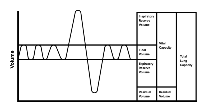
Figure 2 Lung volumes and capacities.
----
Part 4: Gas exchange and regulation
We’ve covered the anatomy and physiology of the respiratory system, and now we’ll analyze the functions in greater detail. As mentioned earlier, aerobic cellular respiration requires oxygen as the final electron acceptor, and this causes a build-up of carbon dioxide that needs to be expelled from the body. This section will cover the mechanism of gas exchange in the lungs, as well as the regulation of gas exchange by the body.
a) Diffusion and differential partial pressure
The primary mechanism of any form of substance transport in the body is diffusion, and there is no exception for respiration. Going back to the earlier chapter about transport, diffusion is the transfer of molecules from a high to low concentration without the consumption of energy. When air enters the lungs and fills the alveoli, many capillaries surround the alveoli to facilitate the transfer of oxygen to the hemoglobin in the blood and carbon dioxide out of the hemoglobin into the lungs for expulsion.
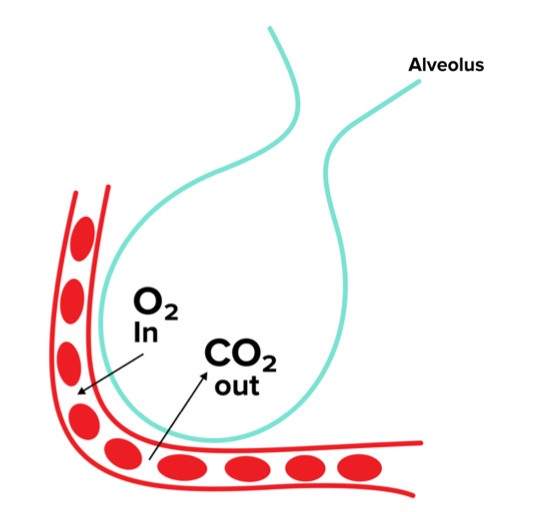
Figure 3 Gas exchange between the alveoli and capillaries.
Deoxygenated blood is mostly saturated with carbon dioxide, and thus the PCO2 in the blood is significantly higher than that of the lungs. This drives carbon dioxide into the lungs. On the other hand, the PO2 in the blood is significantly lower than in the lungs, and thus the pressure gradient pushes oxygen from the lungs into the blood.
b) Henry’s law
How does the gas from the air suddenly diffuse into the liquid blood? Over time, gases in the air that are exposed to liquids equilibrate with the gases in the liquids. Gas molecules from oxygen inhaled in the lungs diffuse into the blood over time. For gas to dissolve into a cell, the gas molecules from the air must dissolve into a liquid. Henry’s law states that the amount of gas that will dissolve into a liquid is proportional to the partial pressure of that gas as well as the solubility of that gas in the specific liquid. For oxygen, the law can be written as such:
In the above equation, [O2] is the concentration of dissolved oxygen, while [PO2] is the partial pressure of oxygen and [SO2] is the solubility of oxygen in a specific liquid. This relationship helps describe how oxygen diffuses into the blood while carbon dioxide diffuses out. As listed above, the partial pressure of oxygen in air is significantly higher than the concentration of dissolved oxygen in the blood, so oxygen diffuses into the blood and vice versa for carbon dioxide.
c) pH and nervous system control
Unless you are focusing on your breathing, it is a subconscious process. As a result, proper regulation of respiration is essential based on cues from inside the body. The main control comes from the respiratory control center in the medulla of the brain stem, which receives signals from the body and regulates respiration. This section will discuss how pH of blood can affect respiration by signaling chemoreceptors in the aorta, arteries, and the brain, as well as how the nervous system responds to these signals.
As discussed earlier, respiration eliminates carbon dioxide from the body, so an absence of respiration leads to a build-up of carbon dioxide. The carbon dioxide combines with the water to form carbonic acid, which further dissociates as an acid into protons and bicarbonate (HCO₃⁻). As you know from chemistry, pH is a measure of the number of protons or hydrogen ions in a solution, and an increase in the concentration of hydrogen ions lowers the pH.
The nervous system is the main party responsible for sensing the change in pH of dissolved gases and adjusting the breathing rate. There are chemoreceptors in the medulla that can tell when the pH is too low due to metabolic acidosis or too high due to metabolic alkalosis. The brain then sends signals to the lungs, leading to a change in respiratory rate to solve these problems in an involuntary manner.
When a patient experiences metabolic acidosis, the equilibrium of the bicarbonate buffer system must shift toward the reactants (leftward) in order to decrease the concentration of H⁺ and increase the pH. As a result, the patient will hyperventilate to “blow off” excess carbon dioxide. Under Le Chatelier’s principle, the bicarbonate present in the blood will then undergo the reverse reaction to become water and carbon dioxide, thus removing H⁺ from the bloodstream.
The opposite is true of a patient experiencing alkalosis. If the pH of the blood is too high and the concentration of H⁺ must be restored, the patient will hypoventilate to retain carbon dioxide. By retaining carbon dioxide, the production of bicarbonate is favored and the concentration of H⁺ increases.
----
Part 5: Overview of the circulatory system
In the previous sections, we talked about how the body provides oxygen to the blood, but now we need to discuss how the blood delivers that oxygen to the rest of the body. Since humans and other animals are such large creatures relative to cell size, the cells have to work together to ensure optimal functioning of the body. The circulatory system helps the cells work together by doing the following: distribution of nutrients, transport of oxygen and carbon dioxide, movement of waste from tissues to kidneys, transport of hormones, regulation of body temperature, and formation of blood clots when necessary. In this and the subsequent sections, we will discuss the basics of the circulatory system you need to know for the MCAT.
a) Components of the circulatory system
The heart is the driving force for blood in the body. Blood that is leaving the heart travels in arteries, and blood that is returning to the heart travels in veins. Arterial blood is high pressure because it has the force of blood being pushed by the heart, and venous blood is low pressure because it is being pulled to the heart. As the arteries get further away from the heart, the pressure decreases and the arteries continuously branch out into arterioles, which have smooth muscle to help control the flow of blood. From arterioles, blood then travels into capillaries, which are tiny vessels where most of the nutrient exchange occurs with body tissues. Each of these types of blood vessels is lined by specialized cells known as endothelial cells. Endothelial cells form a tight seal to prevent the leakage of fluid but can selectively permit the movement of white blood cells or nutrients out of the blood vessel and into the surrounding tissue.
While these capillaries are very small—only big enough for about a single blood cell—there are so many throughout the body that there is ample area for adequate diffusion of gases and materials between the blood and the tissue. From the capillaries, the blood passes to venules, which leads back to veins and then the heart again. Together, these are the main components of the circulatory system.
b) The heart
The heart is a four-chambered organ that is divided in the middle. There are two types of chambers involved in receiving and pumping blood, and they are known as the atria and the ventricles. The atria can be thought of as the receiver of the blood as they are where blood from veins enters the heart before moving on to the ventricles. The ventricles are responsible for the majority of the actual pumping of blood into the arteries.
Since the chambers are separated vertically, the heart is commonly referred to as having a right and left side, with an atrium and ventricle within each. The right atrium is responsible for receiving blood from the body and pumping it to the right ventricle. The right ventricle then pumps the blood to the lungs to obtain oxygen, and then the blood is received again by the left atrium. From the left atrium, blood is pumped to the left ventricle, which pumps the blood to the rest of the body. The flow of blood from the right ventricle through the lungs and back to the left atrium is known as pulmonary circulation, while the blood traveling from the left ventricle through the body and back to the right atrium is known as systemic circulation.
Since the atria and ventricles have different functions, their anatomy is different as well. The atria are mainly responsible for receiving the blood and pumping it only to the ventricles, so they have relatively thin walls and little musculature. The ventricles, on the other hand, have a lot of muscles because they need to pump blood into arteries, and the left ventricle is more muscular than the right because it pumps blood to the entire body instead of just the lungs.
Valves are another important aspect of heart anatomy that ensure a one-directional flow of blood. Since the atrium receives blood and the ventricle pumps blood, there is a significant pressure difference between the two and a great risk of backflow during ventricular contraction. So, there is a need for atrioventricular valves (AV valves) between each atrium and its respective ventricle. The AV valve between the left ventricle and left atrium is called the bicuspid (or mitral) valve, and the valve between the right ventricle and right atrium is the tricuspid valve.
Another set of valves is present between the two ventricles and the arteries in which they pump blood into. These are known as the semilunar valves (SL valves), and they ensure blood does not flow back into the ventricles when they relax. The semilunar valve between the right ventricle and pulmonary artery is the pulmonary valve, and the valve between the left ventricle and the aorta is the aortic valve.
The mechanism and process by which the heart pumps blood is known as the cardiac cycle. It consists of two periods known as systole and diastole. Systole refers to the period when the heart is contracting and actively pumping blood into circulation. Diastole refers to when the heart is relaxed and blood flows into the atria. During diastole, the ventricles are relaxed, and the atria is able to pump blood easily into them. Diastole ends when the ventricles contract, initiating systole, which also closes the AV valves. Systole ends when the ventricles relax and the SL valves close.
Let’s now discuss the heart’s rhythmic control. The heart controls its own rhythm through an electrical signal from the sinoatrial node (SA node) above the right atrium. This means that there is no external signal from the brain to tell the heart to beat—the SA node acts as the pacemaker of the heart. The SA node first sends out an action potential similar to nerve cells, and these are propagated by gap junctions between cardiac muscle cells. This initial action potential travels to both atria, but not the ventricles, causing only the right and left atria to contract. The atrioventricular node (AV node) then propagates the action potential to the ventricles by sending the signal through the bundle of His and into the Purkinje fibers, which spread the impulse evenly throughout both ventricles.
The last concept of the heart we will talk about is cardiac output and how to calculate its value. The heart rate is defined as the number of times the heart beats in a minute, with a typical range of around 45-80 beats per minute (bpm). The stroke volume is defined as the amount of blood pushed through the heart with each beat. Together, these two quantities help us identify the cardiac output, which is the amount of blood pumped through the heart each minute. The equation for cardiac output is as follows:
Cardiac Output = Stroke Volume x Heart Rate
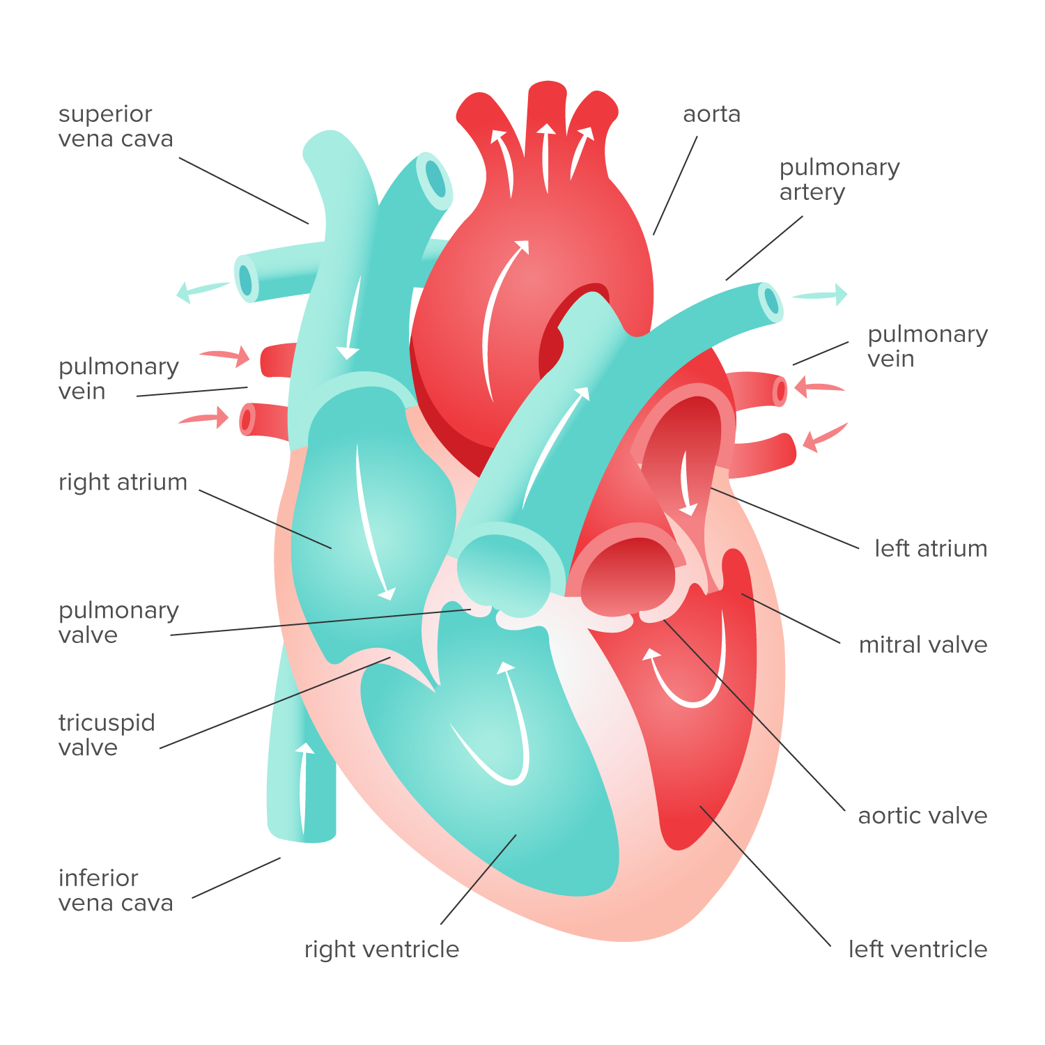
Figure 4 Heart anatomy
----
Part 6: Overview of Blood
The main component of the circulatory system that allows it to fulfill its most basic function is blood. Blood carries materials and gases from one tissue to another, essentially connecting the entire body. If the arteries and veins were subway tracks, the blood can be thought of as the subway car traveling on the tracks. It is important to have a deep understanding of blood for the MCAT, including the components, how it flows, and some of its functions.
a) Blood composition
In the average human, there are about 4-6 liters of blood. There are two main parts of blood: the plasma and the cellular component. Plasma is an aqueous solution that accounts for about 55% of blood volume, and it consists of electrolytes, buffers, sugars, proteins, lipoproteins, gases, and waste products all dissolved in water. The buffers help maintain the blood pH at around 7.4, and a buildup or deficiency of carbon dioxide, as mentioned earlier, can affect this pH.
The quantity of plasma volume and fluid content in the blood is tightly regulated by the kidneys. (For more information on this topic, be sure to refer to our guide on the digestive and excretory systems.) Further, the proteins contained in plasma—including albumin, immunoglobulins, and fibrinogen—assist in maintaining the oncotic pressure of the blood.
The cellular component consists of cells produced in the bone marrow before they are released into the circulatory system. The main components are red blood cells (erythrocytes), white blood cells (leukocytes), and platelets. The volume of blood that contains the erythrocytes is known as hematocrit and is typically around 40-45% in adults. This means leukocytes and platelets take up a very small amount of blood at around only 1%.
Erythrocyte (red blood cell) production is stimulated by a hormone known as erythropoietin. Red blood cells are produced in the red bone marrow of long bones. Erythrocytes are cells that lack a nucleus and organelles. Due to their lack of mitochondria, these specialized cell types must generate energy in the form of ATP using glycolysis. At the end of their lifetime, red blood cells are sent to the spleen (a heavily vascularized accessory organ) for destruction and recycling.
The main purpose of erythrocytes is to transport oxygen from the lungs to the tissues and carbon dioxide from the tissues to the lungs. Their biconcave shape helps with this by creating a larger surface area for gas exchange and allowing them to move more efficiently through capillaries. Additionally, red blood cells contain hemoglobin, which is a protein composed of four subunits. These subunits contain iron-binding cofactors that are specialized for binding gaseous oxygen.
Table 1 Different blood types given genotypes
| IAIA or IAi | IBIB or IBi | IAIB | ii | |
|---|---|---|---|---|
An individual’s blood type is extremely important in clinical settings when donating and receiving blood. For example, a person who has type B+ blood produces anti-A antibodies, and thus if type A blood was transfused, the antibodies would destroy the new blood. Due to the codominant nature of blood types, there are a couple of important types for blood transfusions: AB+ and O-. People that are AB+ do not make antibodies to any of the three antigens (A, B, or Rh), and thus, they are known as universal recipients and can receive any blood type without an adverse reaction. O- patients, on the other hand, produce antibodies for all three antigens and have none of the antigens on their surface, meaning they are universal donors. This also means that if a person with O- blood needs a transfusion, it is extremely difficult to acquire blood for them.
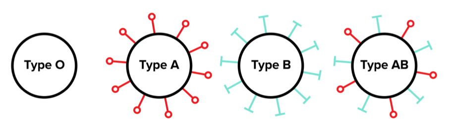
Figure 5 Blood types.
The second type of cell we will discuss is white blood cells, called leukocytes. The role of white blood cells is to fight infection and dispose of debris. There are a few different types of white blood cells, and they function differently. Macrophages and neutrophils move with a crawling mechanism, meaning they can sometimes exit the closed network of arteries and veins and roam around the tissues to fight infections. Other types of white blood cells move through chemotaxis, which, as you may recall, is the movement in response to chemical stimuli, such as waste, toxins, or signals from other white blood cells. Overall, there are 6 types of leukocytes, and they are outlined in the table below.
Table 2 Types of leukocytes
| Cell | Type | Role |
|---|---|---|
| Macrophage | Monocyte | Engulf debris and microorganisms; roaming tissues, chemotaxis |
| B Cell | Lymphocyte | Mature into plasma cell and produce antibodies |
| T Cell | Lymphocyte | Kill virus-infected cells, tumor cells, and reject tissue grafts; control immune response |
| Neutrophil | Granulocyte | Engulf bacteria, roaming tissues, chemotaxis |
| Eosinophil | Granulocyte | Destroy parasites, allergic reactions |
| Basophil | Granulocyte | Store and release histamine; allergic reactions |
The last type of blood cell we will cover is platelets. Platelets are not actually whole cells, but are fragments of larger cells known as megakaryocytes.
Platelets are responsible for initiating clotting and the coagulation cascade during the rupture of a blood vessel. When there is a leak, platelets will react with collagen, thrombin, vitamin K, and calcium to produce a blood clot that prevents further leakage of blood. Once the platelets form the plug, fibrin, a threadlike protein, forms a mesh to hold the clot in place. This mesh dries into what is known as a scab to seal and protect the wound.
b) Blood flow
Understanding blood flow is crucial to the MCAT because it is one of the few ways the test-writers can combine physics with biological concepts. The study of blood flow is known as hemodynamics. The driving force in blood flow is the pressure caused by the heart pumping in one direction. Opposing this force is friction, which is caused by the blood cells against the vessel walls, and the technical term is resistance. Ohm’s Law quantifies the relationship between these variables as ΔP = Q x R, where ΔP is the pressure gradient between the arteries and veins, Q is the blood flow defined by the cardiac output, and R is the resistance. This equation gives us some important conclusions, mainly that blood pressure changes with either cardiac output or resistance (and only those!).
Resistance (R) is modeled in blood vessels through Poiseuille’s Law, which is shown as follows:
Most of these variables are held constant, namely the length (L) of the vessel and the viscosity (η), but the radius, r, changes throughout the body. You may think that the capillaries are significantly smaller, so resistance goes up, but you have to remember there are significantly more capillaries than arteries, and thus the overall radius of capillaries is greater than that of arteries. This means that the pressure actually decreases from arteries to capillaries, and the capillary beds are a source of peripheral vascular resistance.
As the blood continues through the circulatory system, the pressure continues to decrease and is close to zero in the veins right before the heart. Overall, this pressure can be modeled in the figure below.
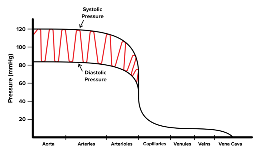
Figure 6 Pressure throughout the circulatory system.
c) Gas exchange by blood
The primary function of red blood cells is to transport oxygen and carbon dioxide to and from the tissues, respectively. These gases, however, are relatively hydrophobic and don’t readily dissolve in the blood. To circumvent this problem, red blood cells contain hemoglobin (Hb), a complex protein with four subunits, and each subunit has a heme group. Heme molecules are a large multi-ring structure that has a single iron atom at its center, and its role is to bind oxygen. Since each hemoglobin has four subunits, each molecule of hemoglobin is capable of binding and carrying four molecules of oxygen.
There are some important properties of hemoglobin to know that make it an excellent oxygen carrier. The way each hemoglobin is structured, the four subunits do not bind oxygen independently. When none of the subunits are bound to oxygen, the hemoglobin assumes what is known as a tense conformation. This tense conformation has a low affinity for oxygen.
When the first subunit binds oxygen, however, the conformation changes to a relaxed state, which has a higher affinity for oxygen. Because of this, hemoglobin is said to bind oxygen in a cooperative fashion. This cooperative binding helps hemoglobin effectively bind oxygen in the lungs and release it in the tissues. In active tissues, oxygen level is very low, and when a single oxygen molecule dissociates from hemoglobin to diffuse into tissues, it aids the rest of the oxygen molecules in dissociating as well.
Another important concept to understand about cooperative binding is the regulation by other factors. Certain conditions within the body help stabilize the tense configuration (remember, no oxygen is bound in this configuration), and it thus reduces the overall affinity of hemoglobin for oxygen. These factors are decreased pH, increased partial pressure of carbon dioxide, and increased temperature. Together, the ability of these factors to stabilize the tense conformation is known as the Bohr effect.
An oxygen-hemoglobin dissociation curve is a common figure to see on the MCAT, so it is important to understand. The tendency of hemoglobin to bind oxygen can be quantified using a % saturation, for which the formula is % sat = (# of bound O2 molecules) / (# of binding sites) x 100%. This allows us to graph the saturation of hemoglobin against the partial pressure of oxygen, as seen in the figure below. You will notice the sigmoidal shape, which is due to the cooperative nature of hemoglobin binding. Additionally, you can see that there is a left-shifted curve and a right-shifted curve. This represents the Bohr effect, with the right-shifted curve causing decreased affinity for oxygen and the left-shifted curve meaning a greater affinity.
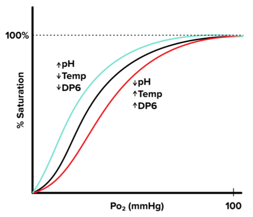
Figure 7 Oxygen-hemoglobin dissociation curve.
While oxygen is the most important gas for bodily function, carbon dioxide is just as important to remove. You may think it binds to hemoglobin the same way that oxygen does to exit the body, but the mechanism is actually entirely different. There are three main ways in which carbon dioxide is transported from the tissues to the lungs. The first way is in the form of carbonic acid. An enzyme called carbonic anhydrase facilitates this process, and it forms a water-soluble molecule that can easily be dissolved in the blood. The second mechanism that accounts for about a fifth of carbon dioxide transport is that the carbon dioxide simply sticks to the hemoglobin. It doesn’t bind at the oxygen-binding sites, but it does bind to various other sites on the protein. The third, and most insignificant method, is by dissolving directly into the blood. Carbon dioxide is more water-soluble than oxygen, so a small portion can be transported directly in the plasma.
----
Part 7: Regulation from other systems
In order for the body to effectively maintain homeostasis, all of the systems must work together, and this integration is important to know for the MCAT because the test-writers like to test your knowledge in unique ways. There are two main systems that play the biggest role in regulating the circulatory system: the nervous system and the endocrine system. This section will discuss some of the ways in which these two systems help regulate different facets of the circulatory system.
a) Nervous system control
While the nervous system doesn’t directly tell the heart to contract, it can regulate the rate of contraction. Without nervous system regulation, the SA node would continuously fire at about 120 beats/min, which is significantly faster than the average human’s heart rate. The lowering of the heart rate results from the parasympathetic nervous system inhibiting the SA node from conducting action potentials that fast. The mechanism through which this occurs is the vagus nerve releasing acetylcholine near the SA node to inhibit depolarization and binding to receptors. The constant inhibition level is known as the vagal tone, and it changes based on the needs of the body.
Another way the nervous system can affect the heart rate is through the sympathetic nervous system in what is commonly known as the “fight-or-flight” response. This works through a series of nerves that innervate the heart and norepinephrine to stimulate muscle contraction. Additionally, epinephrine secreted from the adrenal medulla stimulates muscle in the heart. Together, both of these increase the heart rate as well as the force of contraction.
The last method of regulation by the nervous system is through baroreceptors in the heart. These receptors measure blood pressure and send signals to the central nervous system when the pressure is too high. The CNS then sends signals to the heart to correct this via an increased vagal tone or decreased sympathetic signaling.
b) Endocrine system control
In addition to the nervous system, the endocrine system plays a crucial role in regulating the circulatory system. The main mechanism of regulation is through the release of hormones to alter blood pressure and blood sugar. Since blood plasma is primarily water, hormones that affect fluid retention significantly impact blood pressure. Both aldosterone and antidiuretic hormone (ADH) help retain water. Aldosterone accomplishes this by increasing sodium retention, which increases water reabsorption. ADH, on the other hand, directly increases water absorption in the collecting duct of the nephron.
The endocrine system is also able to decrease water retention with the help of atrial natriuretic peptide (ANP), which decreases sodium absorption and thus decreases water reabsorption. The increase in fluid retention from aldosterone and ADH increases blood pressure, according to Ohm’s Law mentioned earlier, because a greater stroke volume with no change in resistance directly increases pressure.
In addition to blood pressure, insulin and glucagon can affect the sugar content in blood. Insulin is the hormone that facilitates the intake of glucose into cells, thereby reducing blood sugar. The opposite of insulin is glucagon, which facilitates glycogenolysis and gluconeogenesis in liver cells to essentially release glucose into the blood. The back and forth action of these hormones aids in the transport of sugar to different cells in the body and ensures excess sugar is stored in the correct location.
----
Part 8: High-yield terms
Pharynx: tube at the back of the mouth where air passes through; also carries food
Larynx: air tube that contains vocal cords; covered by epiglottis and separates air from food
Trachea: receives air after larynx; commonly referred to as windpipe
Bronchi: carry air from the trachea into the lungs, progressively branching into smaller tubes
Alveoli: microscopic sacs covered with capillaries that are the site of gas exchange between lungs and blood
Surfactant: a soapy substance that coats alveoli to prevent them from collapsing on themselves
Lungs: main organ of the respiratory system; site of gas exchange
Vasodilation: the opening of blood vessels, particularly capillaries, for different purposes
Vasoconstriction: the opposite of vasodilation, the closing of blood vessels
Goblet cells: cells that secrete mucus to help the respiratory system filter out particles and antigens
Epithelial cells: type of cell in the respiratory tract that contains cilia that help sweep the mucus to the pharynx
Diaphragm: primary muscle below the diaphragm that contracts and relaxes to control the flow of air in and out of the lungs
Intercostal muscles: muscles in between the ribs that aid the diaphragm during inhalation
Tidal volume: the volume of air contained in a normal breath
Expiratory reserve volume: the total amount of air that can be exhaled after a passive exhalation
Inspiratory reserve volume: the amount of air that can be inhaled after a passive inhalation
Total lung capacity: the total amount of air that can be contained in the lungs after a full inhalation
Residual volume: the total amount of air that remains in the lungs after a forceful exhalation
Vital capacity: the total amount of air that can be forced out after a full inhalation
Partial pressure: the pressure a specific gas within a mixture of gases exerts; commonly used for pressure of certain gases in air
Acidosis: a condition in the blood where the pH is more acidic, caused by excess carbon dioxide in the blood
Alkalosis: a condition where the blood pH is more basic, caused by a deficiency of carbon dioxide in the blood
Heart: the driving force for blood in the body
Arteries: carry blood away from the heart
Veins: carry blood back towards the heart
Capillaries: blood vessels with very small radii that is the site for nutrient and gas exchange between blood and tissues
Atria: the chambers in the heart that are responsible for receiving blood from veins and pushing it to the ventricles
Ventricles: the chambers in the heart responsible for pumping the blood to the arteries and the rest of the body
Atrioventricular valves: one-way valves that separate the atria and ventricles preventing backflow while ventricles pump blood
Bicuspid valve: specific AV valve separating the left ventricle and left atrium; also called the mitral valve
Tricuspid valve: the specific AV valve between the right atrium and right ventricle
Semilunar valves: the set of one-way valves separating the ventricles from the major arteries they feed into preventing backflow of blood when the ventricles are relaxed
Pulmonary valve: the specific SL valve separating the right ventricle and the pulmonary artery
Aortic valve: the specific SL valve separating the left ventricle and the aorta
Cardiac cycle: the overall mechanism by which the heart pumps blood containing systolic and diastolic periods
Systole: period when the heart is actively contracting and pumping blood into circulation
Diastole: period when the heart is relaxed and blood flows into the atria
SA node: node above the right atrium that sends an electrical signal to both atria to contract and pump blood into the ventricles
AV node: propagates action potential from SA node through the bundle of His and Purkinje fibers to evenly contract both ventricles
Heart rate: defined as the number of times the heart beats per minute; typical range of 45-80 beats per minute in adults
Stroke volume: the amount of blood pushed through the heart with each beat
Cardiac output: defined as the amount of blood pushed through the heart every minute; calculated by multiplying the stroke volume by the heart rate
Plasma: an aqueous solution that composes about 55% of blood; composed of electrolytes, buffers, sugars, proteins, lipoproteins, gases, and waste products all dissolved in water
Erythrocytes: red blood cells; responsible for transporting gases, specifically oxygen
Leukocytes: white blood cells; responsible for fighting diseases in the body
Platelets: type of cell responsible for blood clotting when a vessel ruptures
Hematocrit: the volume of blood containing erythrocytes; typically around 40-45% in adults
Hemoglobin: a protein with 4 subunits that each contain a heme group meant to bind oxygen for effective delivery to the rest of the body
Cooperative binding: the phenomenon by which hemoglobin is more likely to bind oxygen when one of the subunits has already bound oxygen
Bohr effect: the ability of various factors such as temperature and pH to affect the affinity of hemoglobin for oxygen
Vagal tone: the level of inhibition of the SA node by the vagus nerve to lower the heart rate from the 120 bpm the SA node would otherwise produce
----
Part 9: Practice passage and answers
Due to the electrical nature of heart contractility, heart tissue can be analyzed using electrodes. Each electrical impulse from the heart can be recorded on the skin, and the overall pattern makes up what is known as an electrocardiogram (ECG).
A typical ECG gives 2 types of information. The first type is by analyzing the differences between signals. Secondly, it shows us the amount of electrical activity in the heart muscle at a given time. A single heartbeat cycle has 3 wave patterns: a P wave, a QRS complex, and a T wave. These can be visualized in the figure below:
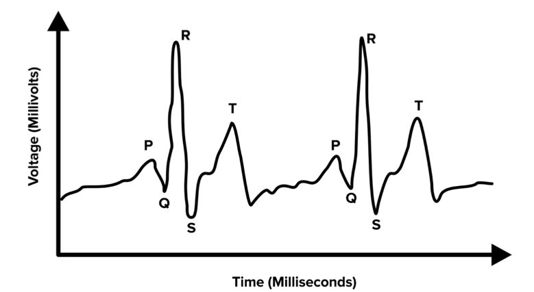
Figure 1
The P wave is caused by depolarization in both atria. The QRS complex is representative of depolarization spreading through the ventricles. The T wave is caused by repolarization of the ventricles.
This passage was created for educational purposes only
Question 1: Which of the following time intervals of an ECG would best represent ventricular diastole?
A) QRS complex
B) T-P region
C) S-T amplitude
D) P Wave
Question 2: A patient with a deficiency in the QRS amplitude would likely have a problem with what?
A) SA node
B) AV node
C) Vagal tone
D) Purkinje fibers
Question 3: If someone were to hold their breath for an extended period of time, which part of an ECG would be most affected?
A) R-R interval
B) QRS amplitude
C) P-Q interval
D) T wave
Question 4: Which of the following would show up on an ECG?
A) Leaky valve
B) Stroke volume
C) Fibrillation
D) High blood pressure
Question 5: A patient with hypothermia would likely experience which of the following in their body?
I. Peripheral vasodilation
II. Piloerection
III. Peripheral vasoconstriction
A) I only
B) I and II only
C) II and III only
D) III only
Question 6: An acetylcholine inhibitor in the heart would NOT affect what heart measurement?
A) Heart rate
B) Hemoglobin saturation
C) Cardiac output
D) Vagal tone
Answer key for passage-based questions
1. The correct answer is B. The question is asking about diastole in the ventricles, which is the period when the ventricles are relaxed. Based on the passage, we see that the T wave is from repolarization of the ventricles, and they are not depolarized until the QRS complex of the subsequent wave. This means that from T-P, the ventricles are relaxed and in diastole. The QRS complex is during ventricular systole (choice A is incorrect), S-T amplitude is irrelevant to time (choice C is incorrect), and the P wave is not the entirety of diastole (choice D is incorrect).
2. The correct answer is D. As mentioned in the passage, the QRS amplitude measures the contraction of the ventricles. The ventricle contraction is spread evenly by the Purkinje fibers, so that is the most likely problematic destination. The SA node would affect the atrial contraction (choice A is incorrect), the AV node simply sends the signal from the SA node to the bundle of His (choice B is incorrect), and the vagal tone impacts the heart rate as a whole (choice C is incorrect).
3. The correct answer is A. This question is combing knowledge about respiration and analysis of the ECG. Holding breath causes a lack of oxygen and thus causes the heart rate to decrease. A heart rate decrease is represented in an ECG by the interval between the R peaks, so an increased R-R interval would mean a decreased heart rate. QRS amplitude means nothing for time (choice B is incorrect), P-Q interval is clinically insignificant (choice C is incorrect), and the T wave is simply the time period of ventricular repolarization (choice D is incorrect).
4. The correct answer is C. An ECG does not tell everything about the heart, but it does tell information about the electrical impulses in the heart. The only choice that has to do with contraction is fibrillation.
5. The correct answer is C. Hypothermia is when the body is extremely cold. The circulatory system controls temperature by dilating and constricting the ventricles to control the amount of blood close to the surface of the skin. A cold body would constrict the vessels, not dilate (choices A and B are incorrect). Piloerection, commonly referred to as goosebumps, is another response to being cold (choice D is incorrect).
6. The correct answer is B. Acetylcholine is the neurotransmitter released by the vagus nerve to suppress the constant pulse by the SA node. If the acetylcholine was inhibited, the vagal tone would decrease (choice D is incorrect), the heart rate would increase (choice A is incorrect), and the cardiac output would also increase (choice C is incorrect), but hemoglobin saturation would remain relatively unchanged.
----
Part 10: Practice standalone questions and answers
Question 1: Which of the following is a correct sequence of blood flow?
A) Pulmonary artery → right atrium → aorta → systemic capillaries
B) Left atrium → arteries → vena cava → systemic circulation
C) Lungs → left atrium → systemic capillaries → right atrium
D) Vena cavae → right ventricle → left atrium → lungs
Question 2: Which of the following contains the vocal cords?
A) Larynx
B) Pharynx
C) Epiglottis
D) Trachea
Question 3: Which blood cell exits blood vessels and roams around tissues to fight disease?
A) Platelets
B) Macrophages
C) B cells
D) Basophils
Question 4: Which blood vessel has the lowest pressure?
A) Arterioles
B) Capillaries
C) Venules
D) Veins
Question 5: A person who is hyperventilating should be expected to have which of the following conditions?
A) Metabolic alkalosis
B) Metabolic acidosis
C) Hyperglycemia
D) Hypoglycemia
Question 6: Which of the following is most likely to occur to someone in a hot environment?
A) Increased secretion from skin glands and constriction of blood vessels
B) Decreased secretion from skin glands and constriction of blood vessels
C) Increased secretion from skin glands and dilation of blood vessels
D) Decreased secretion from skin glands and dilation of blood vessels
Question 7: Which of the following is not involved in protection against disease in the respiratory system?
A) Epiglottis
B) Mucus produced by goblet cells
C) Surfactant in alveoli
D) Cilia on epithelial cells
Answer key for standalone questions
1. The correct answer is C. This question asks about which sequence is correct, and even though the answer choices skip spots in the middle, choice C gives the correct sequence of blood flow.
2. The correct answer is A. The vocal cords are located in the larynx. The pharynx is the tube at the back of the mouth (choice B is incorrect), the epiglottis covers the larynx when swallowing (choice C is incorrect), and the trachea is also known as the windpipe (choice D is incorrect).
3. The correct answer is B. Macrophages are involved in the immune response (choice B is correct). Platelets are blood cells responsible for clotting (choice A is incorrect), B cells produce antibodies (choice C is incorrect), and basophils store and release histamine (choice D is incorrect).
4. The correct answer is D. Looking at the graph of pressure versus the location in the body shown in the content of this guide, we see that pressure continues to decrease from the heart to the veins, where it is close to zero.
5. The correct answer is B. When someone is hyperventilating, the concentration of carbon dioxide in the blood is expected to decrease. Thus, there is less carbon dioxide available to form carbonic acid. Under Le Chatelier’s principle, the concentration of H⁺ will decrease. As a result, the pH of the blood is expected to increase. Hyperventilation is usually observed in response to metabolic acidosis (choice B is correct). Blood sugar is not directly affected by respiration rate (choices C and D are incorrect).
6. The correct answer is C. In a hot environment, people tend to try to dispel heat from the body. One way of doing this is sweating, which is excreted from the skin glands (choices B and D are correct). Another way this is done is by dilating blood vessels to release heat from the blood, making choice C the correct answer.
7. The correct answer is C. While the surfactant coats alveoli, it functions to prevent them from collapsing on themselves. Mucus from goblet cells helps trap particles (choice B is incorrect), and the cilia on epithelial cells sweep the mucus to the pharynx to be swallowed (choice D is incorrect). The epiglottis also protects the larynx from particles while eating (choice A is incorrect).