
Nervous System for the MCAT: Everything You Need to Know
A complete guide to the nervous system on the MCAT, covering key structures, functions, and practice questions to boost your biology score.

(Note: This guide is part of our MCAT Biology series.)
Table of Contents
Part 1: Introduction to the nervous system
Part 2: Divisions of the nervous system
a) Central and peripheral nervous systems
b) Autonomic and somatic nervous systems
c) Sympathetic and parasympathetic nervous systems
Part 3: Microanatomy
a) Anatomy of a neuron
b) Cells of the nervous system
c) The action potential
d) Neural impulses and neurotransmitters
Part 4: Types of Neurons
a) Afferent and efferent neurons
b) Upper and lower motor neurons
Part 5: High-Yield Terms
Part 6: Passage-Based Questions and Answers
Part 7: Standalone Questions and Answers
-----
Part 1: Introduction to the nervous system
From allowing you to perceive your surroundings to remembering your life's most memorable moments, the nervous system can perform some of the most marvelous feats of any organ system. Unfortunately, studies show that many neurological disorders are on the rise. The incidence of Alzheimer's disease, for example, is expected to triple by 2050. Thus, it is more vital than ever that the physicians of tomorrow are equipped with the knowledge needed to care for this growing group of patients.
The information presented in this guide will describe key aspects of the nervous system that are relevant to biology and biochemistry. To better understand the function of the nervous system, be sure to refer to our Psychology and Sociology guides on Psychological Disorders and Behavior and Biology.
-----
Part 2: Divisions of the nervous system
The nervous system is responsible for a variety of functions. Its primary function is to provide control between different body systems. The nervous system serves to integrate information from a variety of body systems, including information about the external environment that is carried into the body, and to coordinate responses that maintain internal homeostasis and proper cellular function.
a) Central and peripheral nervous systems
The nervous system can be divided into two major components. The brain and spinal cord comprise the central nervous system, while nerves and ganglia outside the brain and spinal cord make up the peripheral nervous system.
The outermost part of the brain is the cerebral cortex. This layer merits special attention because it is responsible for many of our higher cognitive functions. The cerebral cortex is rich in the cell bodies, or soma, of neurons. These neurons have long axons that extend through the brain and into the spinal cord.
The cerebral cortex can itself be divided into four major lobes, each with loosely specialized functions.
The frontal lobe governs executive function, initiates voluntary motor movement, and is responsible for producing speech.
The parietal lobe governs spatial processing, proprioception, and somatosensation.
The occipital lobe governs visual processing.
The temporal lobe governs learning, memory, speech perception, and auditory perception. An important language center known as Wernicke’s area is located here.
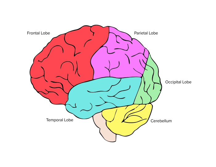
Figure 1 Lobes of the cerebral cortex
Each hemisphere, or side, of the brain is also loosely specialized. The left side of the brain processes sensory information from the right side of the body and is also the primary hemisphere used in performing math and science problems, logical reasoning, and analytical thinking. The right hemisphere of the brain processes sensory information from the left side of the body and is also the primary hemisphere used in spatial awareness, emotional intelligence, intuition, and holistic thinking.
How do these two hemispheres communicate with each other? A structure called the corpus callosum forms a bridge between the left and right hemispheres of the brain. The nerves within the corpus callosum allow sensory information from one side to “cross” to the opposite hemisphere, where it can be processed. Thus, any environmental cues that are sensed by the right side of the body are sent to the left hemisphere of the brain for processing, and vice versa.
b) Autonomic and somatic nervous systems
The peripheral nervous system can be divided into the autonomic and somatic nervous systems. The autonomic nervous system is responsible for regulating the involuntary activities of the body, such as heartbeat, breathing, digestion, and internal body temperature. The autonomic nervous system works closely with a variety of other bodily systems, including the gastrointestinal system and endocrine system. The nervous and endocrine systems integrate closely with each other under feedback control, which results in the stimulation or repression of activation due to the nervous system in response to endocrine products. (For more information on this, be sure to refer to our guide on the endocrine system.)
Conversely, the somatic nervous system is primarily associated with the body’s voluntary movements, including skeletal muscle contraction and relaxation.
c) Sympathetic and parasympathetic nervous systems
The autonomic nervous system can be divided into the parasympathetic and sympathetic nervous systems. The parasympathetic nervous system is often called the “rest and digest system” as it is responsible for digestive processes, such as peristalsis, and sleep-promoting processes, such as slowing the heart rate. The sympathetic nervous system is responsible for the “fight-or-flight” response. The action of the sympathetic nervous system triggers the release of cortisol and epinephrine from the adrenal glands. These hormones and neurotransmitters work together to keep the body alert and prepared to face a stressor, such as by inhibiting digestion, releasing stress hormones, and increasing your heart rate.
Both of these systems are antagonistic to, or oppose, each other.
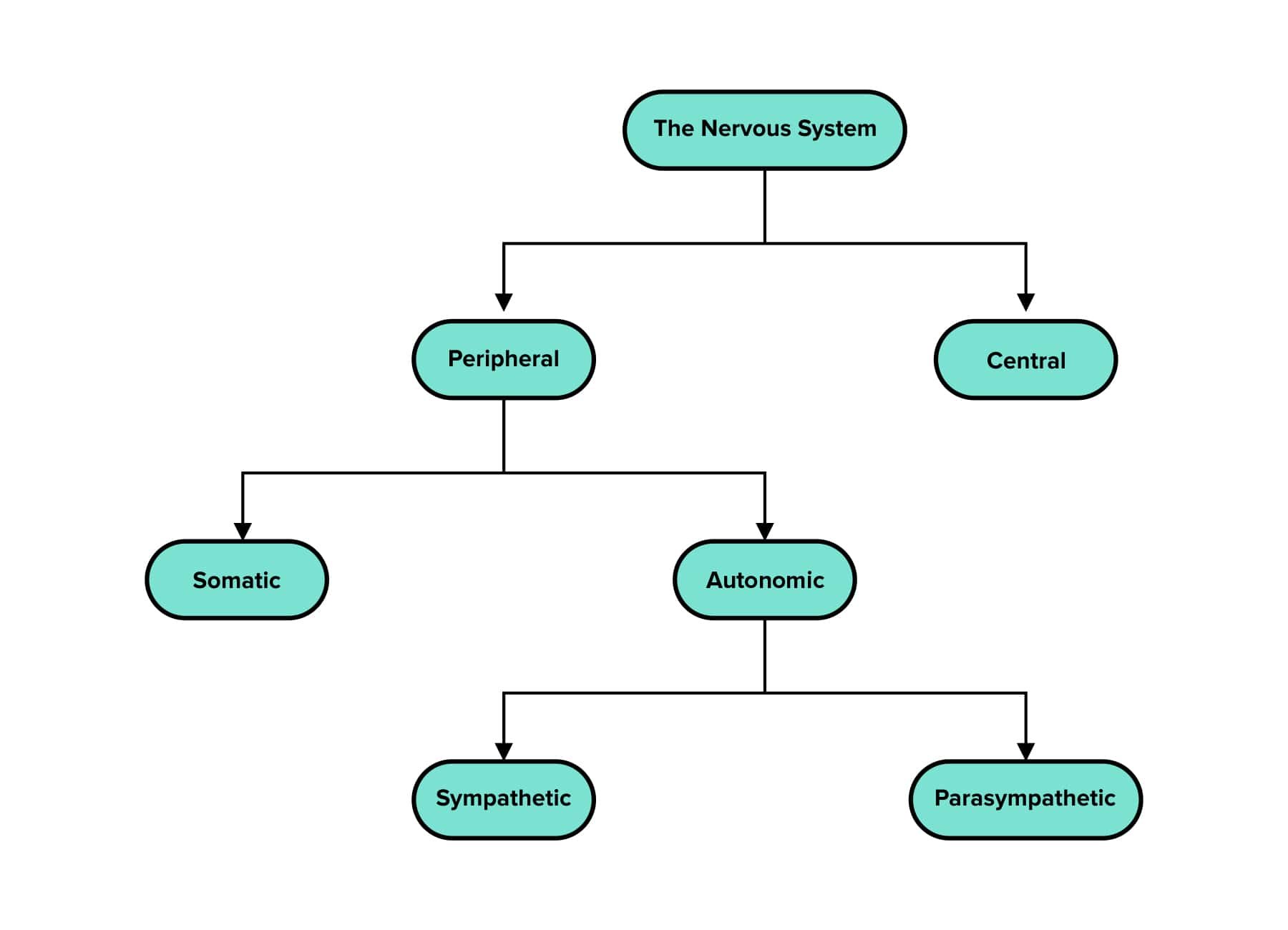
Figure 2 Organization of the nervous system
-----
Part 3: Microanatomy
a) Anatomy of a neuron
The neuron is the workhorse cell of the nervous system. It is characterized by its ability to communicate with other cells using electrical impulses and chemical signals. It contains many different components that you should be familiar with for test day.
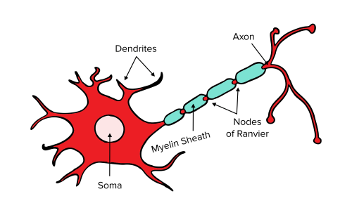
Figure 3 Schematic of a single neuron.
The cell body, or the soma, contains the nucleus, endoplasmic reticulum, and ribosomes of the neuron. Dendrites are outgrowths that branch off from the cell body to receive signals. Axons are long extensions that send signals to other cells via action potentials, which is the electrical impulse neurons use for communication.
A huge variety of helper cells assist in maintaining the structure and function of neurons. To maintain the integrity of the electrical signal and increase conduction speed, oligodendrocytes in the CNS and Schwann cells in the PNS produce myelin—an outgrowth of the helper cell’s plasma membrane—to insulate nerve fibers. Myelin is tightly wrapped around the axon to form sections of insulated axon. The spaces between various sections of myelin sheath are called nodes of Ranvier. These exposed areas of the axon membrane allow for high conduction speeds by allowing the signal to “hop” from node to node, a phenomenon called saltatory conduction.
Signals are passed from neuron to neuron through the synapse, which is made up of the presynaptic neuron’s nerve terminal, a space called the synaptic cleft, and the postsynaptic neuron’s membrane.
Over time, neurons begin to form interconnected pathways that strengthen learned knowledge. This is referred to as synaptic plasticity. Over time, pathways and encoded memories that are used more often will strengthen; pathways that are used less often will weaken. In some cases, entire pathways may die away in a process known as synaptic pruning.
Most neurons in active pathways must remain in place for as long as possible. Neurons generally do not undergo mitosis and instead remain in a senescent state for most of one’s lifetime.
b) Cells of the nervous system
While neurons are the functional cell of the nervous system, they are not the only type of cell present. Neuroglia, or glial cells, are a diverse and important group of cells that support neurons. The table below summarizes these cells, their functions, and if they are specific to the central or peripheral nervous system.
Table 1 Support cells of the nervous system
| Location | Name | Functions |
|---|---|---|
c) The action potential
As we’ve discussed, neurons transmit information through electrical signals, called action potentials, which release neurotransmitters from the presynaptic neuron. The generation of an action potential occurs segmentally and can be divided into four steps: rest, depolarization, repolarization, and hyperpolarization.
When a neuron is at rest (e.g., when an action potential has not yet “fired”), it has a membrane potential of -70 mV. This is due to the presence of charged particles both inside the cell and in the extracellular fluid, including mineral ions and charged proteins. The charges of particles both inside and outside of the cell create an electrical potential across the cellular membrane.
There is a higher concentration of potassium ions inside of the cell compared to the outside of the cell. Potassium leak channels in the membrane allow the positively charged potassium ions to flow out of the cell, which contributes to a net negative charge on the inside of the cell. Conversely, there is a higher concentration of sodium ions outside of the cell compared to the inside of the cell. Sodium leak channels allow positively charged sodium ions to flow into the cell, which partly counters the effects of the outward flow of potassium ions and gives us the -70 mV resting membrane potential. This resting membrane potential is maintained by the sodium-potassium pump, an ATP-dependent enzyme that facilitates the balancing act between sodium and potassium ions. It pumps out three sodium ions for every two potassium ions pumped into the cell.
Inhibitory signals decrease the membrane potential of a neuron through hyperpolarization, or force the membrane potential to become more negative (e.g., less than -70 mV). Conversely, excitatory signals increase the membrane potential of a neuron via depolarization, or force the membrane potential to become more positive (e.g., greater than -70 mV). Once the membrane voltage increases to the threshold value at around -50mV, voltage-gated sodium channels open and allow an influx of sodium ions into one segment of the axon. (Note that reaching the threshold value is an all-or-nothing event: once the threshold value is crossed, the rest of the action potential is guaranteed to fire.) This causes further depolarization until the membrane potential reaches around +35mV to 40mV. This also results in the depolarization of an adjacent segment of the axon.
Once the neuron reaches a membrane potential of +35mV to 40mV, repolarization begins. Now, K+ ions can flow out of the cell as voltage-gated potassium channels open while sodium channels are inactivated. This causes the membrane potential to decrease.
As the potassium ions continue to flow out of the cell, the membrane potential actually becomes lower than the resting potential of -70 mV. This places the neuron into a relative refractory period, during which no stimuli can trigger another action potential.
The sodium-potassium pump also works to restore the resting membrane potential. Recall that for every one molecule of ATP that is hydrolyzed, three Na+ ions are pumped out of the cell and two K+ ions are pumped back in. The cell transitions to the relative refractory period, during which an action potential can occur only if the stimulus is greater than usual.

Figure 4 Voltage changes during an action potential.
Recall that neural impulses are transmitted segmentally in the axon. The influx of sodium ions in one segment during the depolarization phase changes the membrane potential along a short portion of the membrane, thus causing voltage-gated sodium channels in the next segment to open. As the next sodium channels open, depolarization of the membrane causes the cell potential to pass the threshold value. This process continues as the neural impulse propagates along an axon.
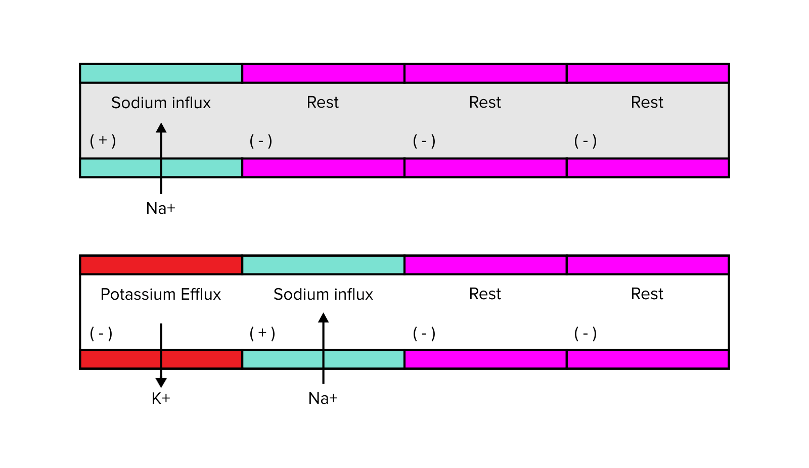
Figure 5 Propagation of an action potential along an axon
d) Neural impulses and neurotransmitters
The propagation of neural impulses can occur electrically or chemically. Electrical synapses are less common and are primarily carried out through gap junctions in cardiac cells. (For more information on the electrical pathways of the heart, be sure to refer to our guide on the respiratory and cardiovascular systems.) Since electrical synapses do not require the flow of chemicals, they are bidirectional and significantly faster than chemical synapses.
Chemical synapses, on the other hand, are the most common way neural impulses are propagated between cells. The exchange occurs at a synapse, or meeting of two neurons. This meeting generally occurs between the terminus, or end, of the axon of a presynaptic neuron and the dendrites of a postsynaptic dendrite.
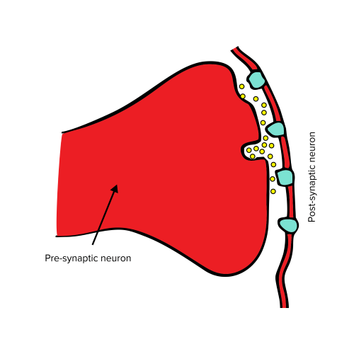
Figure 6 A synaptic cleft.
Chemical synapses are based on the release of neurotransmitters: small-molecule chemical messengers. At the nerve terminal of the presynaptic neuron, the action potential triggers the influx of calcium ions by opening voltage-gated calcium channels. This causes membrane-bound vesicles to release neurotransmitters into the synaptic cleft. The neurotransmitters then bind to receptors on the membrane of the postsynaptic neuron.
The function of each neurotransmitter will directly affect the behavior of the postsynaptic neuron. If the neurotransmitter is excitatory, the postsynaptic neuron will be stimulated to produce its own action potential. If the neurotransmitter is inhibitory, the postsynaptic neuron will be blocked from producing an action potential.
Multiple electrical signals sent into the same synaptic cleft can also sum together to form a larger electrical stimulus. This summation can occur in one of several ways. Temporal summation results from the additive effects of one axon terminal sending repeated, smaller excitatory signals in close succession. Spatial summation results from the additive effects of multiple axon terminals sending multiple smaller excitatory signals to the region around a single post-synaptic neuron.
Neurotransmitters can be removed from the synaptic cleft through one of three possible mechanisms. During reuptake, a neurotransmitter is shuttled into the presynaptic neuron through a transporter on its membrane. In enzymatic degradation, a neurotransmitter is broken down by an enzyme. During diffusion, a neurotransmitter diffuses out of the synaptic cleft and away from the receptors on the postsynaptic neuron. Diffusion is typically the slowest process of neurotransmitter removal, as it depends on the spontaneous flow of molecules down a chemical gradient.
Table 2 Key neurotransmitters of the nervous system
| Name | Regulatory Function | Main Functions | Location | Disorders associated with deficit |
|---|---|---|---|---|
-----
Part 4: Types of neurons
a) Afferent and efferent neurons
Afferent neurons, or sensory neurons, send signals to the brain and spinal cord from sensory receptors. Efferent neurons, or motor neurons, send motor information from the brain and spinal cord to effectors, such as muscles. (It may be helpful to use this mnemonic: afferent nerves arrive at the brain, while efferent nerves exit.)
Interneurons are neurons that function as intermediaries between afferent and efferent neurons. Interneurons are most notable for their role in reflexes. An afferent neuron may send a signal to an interneuron in the spinal cord, which can quickly send a signal to an efferent neuron; this is a process that can occur in the spinal cord without the intervention of the brain.
The most common example of this is the withdrawal reflex. If you were to accidentally place your hand on a hot stove, afferent neurons would send the sensory signals of temperature and pain to interneurons in your spinal cord that are also linked to efferent neurons. The efferent neurons are able to coordinate multiple muscles so that you retract your hand quickly rather than waiting for the signals to reach the brain. This is a type of polysynaptic reflex arc as it involves at least one interneuron linking afferent and efferent neurons. Monosynaptic reflex arcs, on the other hand, consist only of an afferent and efferent neuron with one synapse. A common example of this is the knee-jerk reflex, during which afferent neurons in the knee synapse with an efferent neuron in the spinal cord. This stimulates muscles in the leg, causing it to extend.
Although reflexes are fast and efficient at removing us from sources of danger, the motions they cause are often imprecise and broad.
b) Upper and lower motor neurons
Motor neurons can be further classified into two main categories: upper motor neurons and lower motor neurons. In a nutshell, upper motor neurons in the cerebral cortex transmit information to lower motor neurons in the spinal cord. Lower motor neurons then meet the skeletal muscle at neuromuscular junctions, which stimulates muscular contraction.
Defects of upper motor neurons are associated with four primary abnormalities, or signs. These signs can be thought of as a result of “overstimulation” of the lower motor neuron: a defect in the upper motor neuron causes an abnormally large number of stimulatory signals to be sent to the lower motor neuron.
Hyperreflexia: Hypersensitive receptors in muscles result in greater stretch reflexes
Hypertonia: Increased muscle tension and tightness inhibits muscles from being able to stretch fully
Clonus: repeating muscular contractions and relaxations
Extensor plantar response: Stimulation of the bottom of the foot causes the toes to extend up when normally they should exhibit a downward response
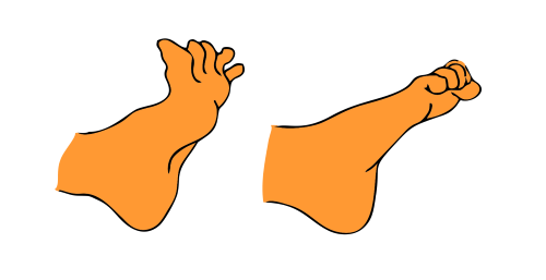
Figure 7 Extensor plantar response (left); normal response (right)
Defects of lower motor neurons are associated with four primary abnormalities, or signs. These can be thought of as the result of a lack of receptivity by the lower motor neuron. Defects in the lower motor neuron weaken the resulting signal to the muscle it meets.
Hyporeflexia: Diminished receptor sensitivity in muscles resulting in decreased stretch reflexes
Hypotonia: Decreased muscle tension and tightness, which inhibits muscles from being able to stretch fully
Fasciculations: Spontaneous, involuntary muscle twitching
Muscular atrophy: Degradation of skeletal muscle
-----
Part 5: High-yield terms
Corpus callosum: a nerve tract that forms a bridge between the left and right hemispheres of the brain
Parasympathetic nervous system: responsible for the “rest and digest” response
Sympathetic nervous system: responsible for the “fight-or-flight” response
Dendrites: branch off from the neuronal cell body to receive signals
Axons: long extensions of the neuron that send signals to other cells
Neuroglia: a diverse and important group of cells that support neurons
Sodium-potassium pump: hydrolyzes 1 ATP to pump 3 sodium ions out of the cell and 2 potassium ions into the cell
Absolute refractory period: period during which no stimuli can trigger another action potential
Relative refractory period: period during which an action potential can occur only if the excitatory stimulus is greater than usual
Synapse: meeting of two neurons that generally occurs between the end of a presynaptic axon and postsynaptic dendrite
Neurotransmitters: small-molecule chemical messengers that pass between neurons at a synapse
Reuptake: process of neurotransmitter removal in which a neurotransmitter is shuttled into the presynaptic neuron through a transporter on its membrane
Afferent neurons: sensory neurons
Efferent neurons: motor neurons
Polysynaptic reflex arc: reflex arc that involves at least one interneuron linking afferent and efferent neurons
Monosynaptic reflex arcs: reflex arcs with only one synapse between an afferent and efferent neuron
Upper motor neuron: efferent neurons that synapse from the brain into the spinal cord
Lower motor neuron: efferent neurons that synapse from the spinal cord onto the neuromuscular junction
-----
Part 6: Passage-based questions and answers
Demyelinating diseases cause destruction of the myelin sheath. Demyelination can be the result of an inflammatory process, viral infection, acquired metabolic derangement, or cardiovascular disruption. Diseases that can cause inflammatory demyelination of the CNS include multiple sclerosis (MS), acute disseminated encephalomyelitis (ADEM), and acute hemorrhagic leukoencephalitis. Demyelinating diseases can also cause acute respiratory failure when the spinal cord is affected.
A 17-year-old Greek female was hospitalized in the ICU due to acute respiratory failure requiring mechanical ventilation. The patient had a history of febrile disease one month previous to the visit, which included an acute onset of paraplegia, diplopia, progressive one-sided arm weakness, and dyspnea. A demyelinating peripheral nervous system (PNS) disease was determined to be the underlying condition.
An MRI of the brain revealed the presence of lesions involving the optic nerve, basal ganglia cerebellum, pons, and medulla oblongata. There was also extended involvement along the spinal cord. Further, blood work revealed elevated counts of white blood cells within the cerebrospinal fluid.
The patient required mechanical ventilation for two months. Then she was transferred to a rehabilitation center. Three years later she remains paraplegic. Since then, she has not suffered any other demyelination attack.
Question 1: Based on the information in the passage, which of the following is a possible effect of the patient’s underlying condition?
A) Decreased synaptic speed
B) Decreased conduction speed of electrical signals
C) Increased sensitivity in extremities
D) Decreased rates of cognition
Question 2: The cellular structures that received damage in this patient are most directly responsible for which of the following functions?
A) Sending signals to other cells
B) Receiving signals from other cells
C) Synthesizing protein
D) Housing genetic material
Question 3: The patient’s arm weakness is most likely due to a defect in which of the following structures?
A) Afferent neurons in the central nervous system
B) Efferent neurons in the peripheral nervous system
C) Sensory neurons in the peripheral nervous system
D) Interneurons in the spinal cord
Question 4: According to the information presented in the passage, damage to which of the following structures would result in the physical symptoms presented by this patient?
A) Schwann cells
B) Oligodendrocytes
C) Upper motor neurons
D) Lower motor neurons
Question 5: Which of the following accurately describes the relative refractory period after an action potential?
A) An action potential is unable to be generated again by any stimulus
B) An action potential is able to be generated again by a weaker stimulus
C) An action potential is able to be generated again by a stronger than usual stimulus
D) An action potential is able to be generated again by a normal stimulus, but it becomes more energetically costly
Answer key for passage-based questions
Answer choice B is correct. The passage states that the patient has an underlying demyelinating central nervous system disease. Myelin insulates nerve fibers, which increases the conduction speed of electrical signals along the length of axons. Thus, there would be a conduction speed along the length of the axon (choice B is correct). Since demyelination does not affect nerve terminals, the speed of synapses is not affected by demyelination (choice A is incorrect). While cognition and sensitivity may be affected, there is not enough information to presume this would occur (choices C and D are incorrect).
Answer choice A is correct. Axons are long outgrowths from the neuron that send signals to other cells (choice A is correct). Dendrites are shorter outgrowths that serve to receive signals (choice B is incorrect). The soma, or cell body, is the site of the nucleus, endoplasmic reticulum, and the ribosomes, which synthesize proteins (choices C and D are incorrect).
Answer choice B is correct. Efferent neurons carry information away from the brain to stimulate muscle contraction and movement. The peripheral nervous system includes all neurons that are not in the brain or spinal cord, including nerves that innervate the arm (choice B is correct). Afferent neurons carry sensory information into the brain (choices A and C are incorrect). Interneurons are most often implicated in reflex arcs and are not relevant here (choice D is incorrect).
Answer choice A is correct. This patient has suffered a demyelinating disease, which possibly targets glial (helper) cells that produce myelin. The patient’s one-sided arm weakness indicates that her symptoms are indicative of a PNS defect rather than a CNS defect. Oligodendrocytes myelinate nerve fibers in the central nervous system (choice B is incorrect). Schwann cells myelinate nerve fibers in the peripheral nervous system (choice A is correct). While the patient’s upper and lower motor neurons may also have been damaged, there is no explicit evidence that the neurons themselves have been targeted (choices C and D are incorrect).
Answer choice C is correct. During the relative refractory period, an action potential can only be generated again by a stronger than usual stimulus (choices B and D are incorrect). During the absolute refractory period, an action potential is unable to be generated again by any stimulus until the cell is back to its resting membrane potential (choice A is incorrect).
-----
Part 7: Standalone questions and answers
Question 1: Which of the following accurately describes the sodium-potassium pump?
A) It is an ATP-dependent enzyme that pumps out two sodium ions for every three potassium ions brought into the cell
B) It is an ATP-independent enzyme that pumps out three sodium ions for every two potassium ions brought into the cell
C) It is an ATP-dependent enzyme that pumps out three sodium ions for every two potassium ions brought into the cell
D) It is an ATP-independent enzyme that brings in three sodium ions for every two potassium ions pumped out of the cell
Question 2: Which of the following molecules is an inhibitory neurotransmitter in the central nervous system?
A) GABA
B) Glycine
C) Glutamate
D) Epinephrine
Question 3: A patient with an upper motor neuron defect may exhibit which of the following symptoms?
A) Hypotonia
B) Fasciculations
C) Muscular atrophy
D) Hypertonia
Question 4: What is the threshold value for a neuron to initiate an action potential?
A) +35 mV
B) -50 mV
C) -70 mV
D) 0 mV
Question 5: Which of the following is NOT a mechanism by which a neurotransmitter can be removed from the synaptic cleft?
A) Enzymatic degradation
B) Reuptake
C) Lysosomal digestion
D) Diffusion
Question 6: The influx of calcium ions into the neuron most directly causes which of the following to occur?
A) Hyperpolarization of the neuron
B) Release of neurotransmitters into the synaptic cleft
C) Sequestration of neurotransmitters into storage vesicles
D) The repolarization of the neuron
Answer key for standalone questions
Answer choice C is correct. The sodium-potassium pump is an ATP-dependent enzyme (choices B and D are incorrect). It pumps out three sodium ions for every two potassium ions brought into the cell (choice C is correct).
Answer choice A is correct. GABA is the primary inhibitory neurotransmitter of the central nervous system (choice A is correct). Glycine is the primary inhibitory neurotransmitter of the peripheral nervous system (choice B is incorrect). Glutamate is the primary excitatory neurotransmitter of the central nervous system (choice C is incorrect). Epinephrine is involved with alertness, sympathetic nervous system responses, and memory formation (choice D is incorrect).
Answer choice D is correct. Hypertonia is one of the four primary signs of upper motor neuron defects (choice D is correct). Hypotonia, fasciculations, and muscular atrophy are signs of lower motor neuron defects (choices A, B, and C are incorrect).
Answer choice B is correct. -50 mV is the threshold value at which an action potential is generated (choice B is correct). Repolarization is initiated when the cell membrane reaches +35 mV (choice A is incorrect). The resting membrane potential of the cell is equal to -70 mV (choice C is incorrect). There is no special event that occurs when the membrane reaches 0 mV (choice D is incorrect).
Answer choice C is correct. While lysosomes are responsible for breakdown of particles inside the cell, neurotransmitters that need to be removed from the synaptic cleft are found outside of neurons in the synapse. Thus, lysosomes would not be able to digest neurotransmitters (choice C is correct).
Answer choice B is correct. Action potentials trigger the influx of calcium ions that cause the release of neurotransmitters into the synaptic cleft (choice B is correct). The neurotransmitters would be stored in vesicles prior to this event (choice C is incorrect). Hyperpolarization and repolarization are due to the efflux of potassium ions (choices A and D are incorrect).