
Behavior and Biology for the MCAT: Everything You Need to Know
Learn high-yield behavior and biology topics for the MCAT, including learning, cognition, social processes, and biology, with practice questions.

(Note: This guide is part of our MCAT Psychology and Sociology series .)
Part 1: Introduction to biology and behavior
Part 2: Anatomy of the nervous system
a) Divisions of the nervous system
b) Neurotransmitters
c) Regions of the brain
Part 3: Reflexes and Innate Behavior
Part 4: Methods Used to Study the Brain
Part 5: High-Yield Terms
Part 6: Passage-Based Questions and Answers
Part 7: Standalone questions and answers
-----
Part 1: Introduction to biology and behavior
Behavior refers to any and all of the actions our bodies perform: whether they are intentional, such as solving a mathematical equation, or unintentional, such as a knee-jerk reflex. Behavior can also include personality, cognition, and decision-making.
Our behavior is influenced by a complex interplay between our environment, our genes, and a variety of biological systems. Of these biological systems, perhaps the most important is the nervous system. In this guide, we'll begin to introduce the role of the nervous and endocrine systems in shaping our behavior and interactions with the environment. What are the specific structures and pathways that are important to our behavior? And what happens to our behavior when these biological systems are disrupted or damaged?
The information presented in this guide will describe key aspects of the nervous system that are relevant to behavior. To better understand the nervous system in its entirety, be sure to refer to our guide on the nervous system.
Throughout this guide, several important keywords will be given in bold. You are encouraged to try creating your own definitions and examples to better aid your understanding. Similarly, we encourage you to sketch diagrams and annotate them with your own knowledge to deepen your understanding of different concepts. At the end of this guide, there are also MCAT-style practice questions that will test your knowledge of this material.
Receive Your Free MCAT Question of the Day
Get every last bit of practice in before test day with a free MCAT question delivered straight to your inbox daily.
-----
Part 2: Anatomy of the nervous system
a) Divisions of the nervous system
The nervous system can be divided into the central nervous system (the brain and spinal cord) and the peripheral nervous system. The names of these divisions of the nervous system are intuitive; they match their location and function. After all, the brain and spinal cord are responsible for processing information and coordinating tasks with the peripheral nervous system, which includes all sensory neurons and nerves that move the muscles.
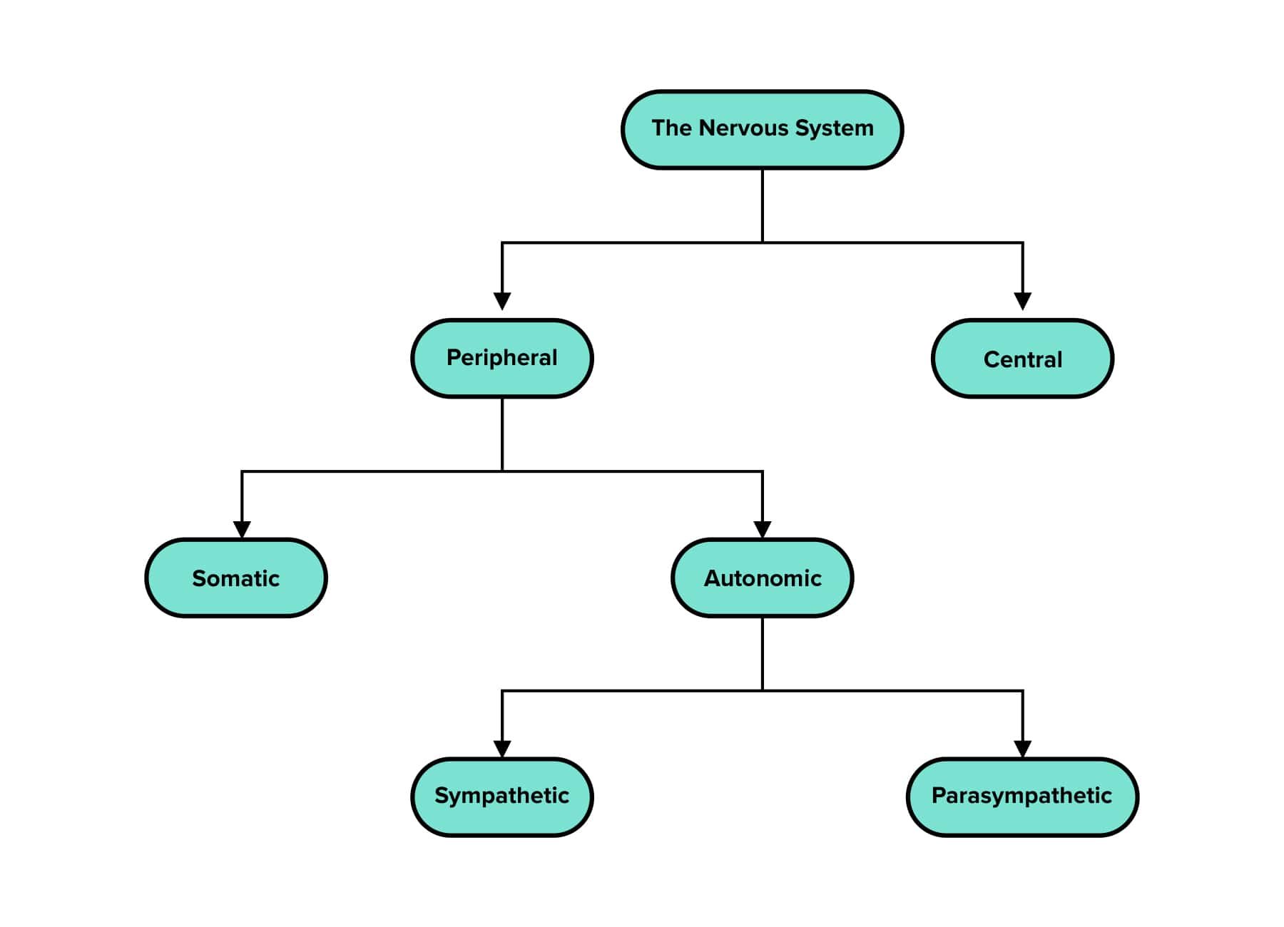
Figure 1 Divisions of the nervous system.
The peripheral nervous system and central nervous system communicate with each other through the peripheral nervous system’s efferent and afferent nerves. Afferent nerves are sensory nerves that ascend the spinal cord and carry sensory information from the environment into the brain. Efferent nerves are motor nerves that exit the brain and descend the spinal cord, relaying commands from the brain to our muscles. (It may be helpful to use this mnemonic: afferent nerves arrive at the brain, while efferent nerves exit.)
Afferent and efferent fibers are organized in the spinal cord and generally occupy different regions. The dorsal roots (back-facing side) of the spinal cord contain bundles of afferent (sensory) fibers. The ventral roots (belly-facing side) of the spinal cord contain bundles of efferent (motor) fibers.
The peripheral nervous system can be further divided into the somatic and autonomic nervous systems. The somatic nervous system governs voluntary movements and thus innervates the skeletal muscle. In contrast, the autonomic nervous system governs automatic or involuntary movements. This includes nervous system control of the digestive system, various glands, and smooth muscle.
The autonomic nervous system is further divided into the sympathetic (“fight-or-flight”) and parasympathetic (“rest-and-digest”) nervous systems. The sympathetic nervous system is designed to respond to stressors in the environment, and the parasympathetic nervous system is designed to help us rest. The sympathetic nervous system expends energy, while the parasympathetic nervous system conserves energy.
In response to a stressor, the sympathetic nervous system will cause:
inhibition of saliva production
dilation of the pupils: to take in as much light as possible to better assess the dangerous situation
accelerated heart rate: to transport oxygen to the muscles
slowed digestion: so blood can be diverted to the muscles
airway dilation: to provide more oxygen with each inhalation
increased glycogenolysis in the liver and increased blood glucose levels: to provide energy
Activation of the parasympathetic nervous system would cause the opposite effects. This includes the diversion of blood to the digestive tract so nutrients may be absorbed and glycogenesis in the liver is increased. (For more information on glycogenolysis and glycogenesis, refer to our guide on carbohydrate metabolism.)
When presented with a stressor or dangerous situation, the sympathetic nervous system is activated. The adrenal cortex releases cortisol while the adrenal medulla releases epinephrine: an important hormone and neurotransmitter, respectively, implicated in the stress response. Additionally, a cognitive appraisal of the situation is performed. During this appraisal, the ability to resolve the stressor or situation is assessed and likely outcomes are determined.
b) Neurotransmitters
Neurotransmitters are chemical messengers that transmit messages from a neuron cell to a target cell. They can be classified based on function (e.g., mood regulation), effect on the cell membrane potential (e.g., inhibitory, excitatory, or both), or locus of action (e.g., central or peripheral nervous system). For example, acetylcholine is the key excitatory neurotransmitter of the skeletal system and regulates movement.
Table 1 Summary Table of Important Neurotransmitters on the MCAT
| Name | Function | Main Functions | Location | Disorders associated with deficit |
|---|---|---|---|---|
You should remember how neurotransmitters function in relation to membrane potential. Neurotransmitters exit via exocytosis from the presynaptic terminal, travel across the synaptic cleft, and then bind to receptors on the target cell where they induce changes in the neuronal membrane's permeability to ions.
As noted, deficits in any of these neurotransmitters may cause disorder or illness. To learn more about this, be sure to refer to our guide on psychological disorders.
c) Regions of the brain
The brain is roughly composed of three main areas: 1) the hindbrain, 2) midbrain, and 3) forebrain. In humans, the brainstem is composed of regions from the midbrain and hindbrain. The functions carried out by these areas of the brain correspond with their evolutionary age: the ancient hindbrain governs basic survival mechanisms (including breathing), while the younger forebrain governs complex functions (such as interpreting language). As a result, while individuals can generally live without a forebrain, they would die without a midbrain or hindbrain.
Table 2 Key Forebrain, Midbrain and Hindbrain functions
| Substructure | Derived from | Main Functions |
|---|---|---|
The outermost part of the cerebrum is the cerebral cortex. This layer merits special attention because it is responsible for many of our higher cognitive functions. The cerebral cortex is rich with the cell bodies, or soma, of neurons. These neurons have long axons that extend through the brain and into the spinal cord.
The cerebral cortex can itself be divided into four major lobes, each with loosely specialized functions.
The frontal lobe governs executive function, initiates voluntary motor movement, and is responsible for producing speech.
The parietal lobe governs spatial processing, proprioception, and somatosensation.
The occipital lobe governs visual processing.
The temporal lobe governs learning, memory, speech perception, and auditory perception. An important language center known as Wernicke’s area is located here.

Figure 2 The four major lobes of the cerebral cortex.
Note that the left and right hemispheres of the cerebral cortex are connected by a bundle of nerves called the corpus callosum. The corpus callosum is essential to ensuring that two sides of the brain can communicate with one another. It also allows the sending of sensory information from one side of the body to the opposite side of the brain for processing. Thus, any environmental cues that are sensed by the right side of the body are sent to the left hemisphere of the brain for processing. One exception to this rule is the perception of smell or odor; in olfaction, sensory signals are sent directly into the brain by cranial nerves without crossing to the opposite hemisphere.
Wernicke’s area and Broca’s area are two key regions of the brain that are necessary for language skills.
Wernicke’s area is located on the left side of the temporal lobe and is necessary for understanding speech. Wernicke’s aphasia can result from traumatic injury to this area. Patients with Wernicke’s aphasia find it difficult to understand written and spoken language but speak a nonsensical stream of words with the fluidity and intonations of regular speech.
Broca’s area is located on the left side of the frontal lobe and is responsible for speech production. Broca’s aphasia can result from traumatic injury to this area. Patients with Broca’s aphasia can understand written and spoken language well but may be unable to form coherent words or speech.
-----
Part 3: Reflexes and innate behavior
Reflexes are behaviors that do not need to be taught or learned. They are the result of neuronal pathways that have been preserved and strengthened over many years of evolution.
The neonatal reflexes are naturally present in newborn infants and gradually disappear as the child matures. These reflexes are normal and expected. However, the presence of a neonatal reflex well after infancy could indicate a neurological problem.
The Babinski reflex is present in children from ages 0-2 years old. It is stimulated by dragging a pointed object across the sole of the foot. A normal response is plantar extension, in which the toes splay or extend outwards. In adults, the reflex disappears and the toes curl inward (flex) instead.
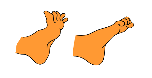
Figure 3 Plantar extension and flexion.
The grasping reflex is present in children from ages 0-6 months old. It is stimulated by placing an object in the infant’s hand. In response, the infant will tightly clasp its hand around the object.
The rooting reflex is present in children from ages 0-4 months old. It is stimulated by stroking the cheek. In response, the infant will turn the head to the same side as the cheek that is stimulated. This reflex is thought to be an evolutionary adaptation to suckling; by turning the head, the infant is able to find the mother’s nipple and attach itself.
The sucking reflex is present in children from ages 0-7 months old. It is stimulated by placing an object in the infant’s mouth or stroking the roof of the mouth. This results in the infant suckling or sucking on the object that was placed in the mouth. This is an unconscious reflex that becomes a conscious response later in infancy and maturity.
The Moro reflex is present in children from ages 0-6 months old. In response to a startling sound or abrupt change in the infant’s position, the infant will rapidly reach its arms outward with outspread hands and then draw its arms closer together.
The stepping reflex is present in children from ages 0-2 months old. When the infant is held upright such that the feet can touch the floor, it will take step-like motions on the ground. This is a developmental precursor to walking, which is usually attained around 1 year of age.
The tonic neck reflex is present in children from ages 0-7 months old. When the infant is placed lying down with the head turned to one side, the arms move in response. The arm on the same side the infant’s head is turned to will extend while the opposite arm flexes. Due to the resemblance of this posture to a fencing stance, this reflex is also known as the fencing reflex.
-----
Part 4: Methods used to study the brain
It’s important to remember that all behavior originates from the neural connections that are formed in the brain. So, to understand a particular behavior, researchers must be able to non-invasively study the brain.
There are several popular imaging techniques you should be familiar with for the MCAT.
A computerized tomography (CT) scan is formed by composing several X-ray images together. After a patient is placed into a CT scanner, multiple X-ray images are taken from multiple angles. These images can be stitched together to form a rough three-dimensional image of the inside of the patient’s body. While this form of imaging is fast and relatively cheap, there are limitations. Importantly, CT lacks the ability to resolve fine detail and accurately model the soft tissues of the brain.
A positron emission tomography (PET) scan may be considered an “improvement” on a CT scan. While the imaging setup is exactly the same, the patient must also be injected with a radioactive tracer beforehand. The type of radioactive tracer that is injected may be tailored to a specific cell activity that must be detected. For instance, a tracer may be used that behaves like glucose and is thus consumed by cells with high metabolic rates. The radioactive tracer releases gamma rays with high energy that can be detected by specialized cameras. (For more information on gamma decay and subatomic particles, be sure to refer to our guide on atomic and nuclear physics.)
While CT and PET scans are limited in their ability to capture changes in activity in real-time, magnetic resonance imaging (MRI) and (functional magnetic resonance imaging) fMRI scans offer high-resolution images of both soft and hard tissues within the body. These scanners use highly powerful electromagnets to create magnetic fields around the body, which interact with the nuclei of atoms within the body. The energy released by these interactions can be detected by a sensor, which creates highly detailed images in nearly real-time.
MRI scans are used to image soft tissues at rest. fMRI is used to measure blood flow through certain areas of the body, under the assumption that tissues with a high metabolic rate require higher levels of blood flow. Due to its high temporal and spatial resolution and the ability to detect changes in real-time, fMRI is the preferred method of choice for imaging the activity of the brain as patients perform tasks.
Lastly, electroencephalograms (EEG) can be used to study the electrical activity of the brain. For more information on reading and interpreting an EEG, be sure to refer to our guide on consciousness, sensation, and perception.
-----
Part 5: High-yield terms
Afferent nerves (also known as sensory nerves): nerves that relay sensory information from receptors in skin or muscle by ascending the spinal cord
Autonomic nervous system: governs our automatic or involuntary movements (movements that occur without our conscious control) and innervates our digestive system, various glands, and smooth muscle
Brainstem: the hindbrain and midbrain
Central nervous system: the brain and spinal cord
Corpus callosum: a bundle of nerves that connects the left and right hemispheres of the brain
Dorsal roots: the back-facing side of the spinal cord that contains bundles of sensory, or afferent, fibers
Efferent nerves: (also known as motor nerves) exit (down) the spinal cord to relay motor information or commands from the brain to our muscles
Grasping reflex: a neonatal reflex in which an infant tightly clasps its hand around an object placed in the palm
Peripheral nervous system: all nervous tissue, excluding the brain and spinal cord
Neonatal reflex: an innate, normal, and expected response to a stimulus; appears as a newborn and disappears sometime during infancy
Neurotransmitters: chemical messengers that transmit messages from a neuron cell to a target cell
Parasympathetic nervous system: a subdivision of the autonomic nervous system that conserves energy and promotes rest through “rest-and-digest” responses
Rooting reflex: a neonatal reflex where an infant turns their cheek toward the side their cheek is touched in the hopes of finding the mother’s nipple
Somatic nervous system: a subdivision of the nervous system as a whole that governs our voluntary movements and innervates skeletal muscle; contrasted with the autonomic nervous system
Suckling reflex: a neonatal reflex where an infant sucks on an object placed in the mouth
Sympathetic nervous system: a subdivision of the autonomic nervous system that governs physiological responses to stressors or “fight-or-flight” responses
Ventral roots: the belly-facing side of the spinal cord that contains bundles of motor or efferent fibers
-----
Part 6: Passage-based questions and answers
Experiences such as “seeing in the mind’s eye,” or “hearing in the head,” refer to quasi-perceptual mentation that resembles vivid perception in the absence of any external stimuli. Among various types of mental imagery, musical imagery appears to be surprisingly accurate. Little is known about how different regions of the brain are activated and subsequently interact in networks.
Researchers investigating this phenomenon had mapped and compared cortical activations and functional networks when producing musical imagery. Silent music visualization was used to guide experienced musicians to repeatedly imagine an eight-minute symphony in the absence of external auditory input. The visualization served to inform subjects of the music content and control the timing for participants’ imagery experience. Participants were also encouraged to imagine the music in a similar way as they listened to it.
The fMRI data acquired with this paradigm were used to map cortical activations and functional networks that resulted. When a participant with musical training listened to an eight-minute music piece eight times during the fMRI acquisition, activated cortical areas were mapped.
The activated areas were found to be mostly confined to the auditory cortex, as shown in Figure 1.
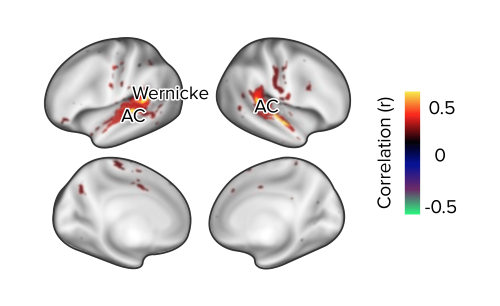
Figure 1 Musical perception and cortical activation measured using fMRI.
Brain activity appeared to be slightly stronger in the right hemisphere than the left hemisphere, with strong activity demonstrated in Wernicke’s area. Using this information, researchers were able to further investigate how the brain engages cortical processes to support musical imagery in the absence of any auditory input.
Question 1: Which of the following accurately describes the imaging method used by the researchers?
A) Radioactive tracer molecules are injected into the patient
B) An electrode cap is placed onto the patient’s scalp
C) Blood flow to different parts of the brain are measured
D) Levels of glucose in the brain are measured
Question 2: The hemisphere in which Wernicke’s area is located is most associated with which of the following cognitive functions?
A) Right-sided odor perception
B) Right-hand control
C) Spatial awareness
D) Face recognition
Question 3: The auditory cortex belongs to which of the following structures?
A) Brainstem
B) Hypothalamus
C) Occipital lobe
D) Temporal lobe
Question 4: Suppose that instead of a symphony, the participants listened to a soundtrack of sudden, loud noises such as car horns and alarms. Which of the following physiological changes would most likely occur?
A) Slowed secretion of digestive juices
B) Increased rate of glycogen storage
C) Constricted pupils
D) Slowed heart rate
Question 5: Which of the following statements accurately describes the neurons that initially receive auditory signals?
A) Motor neurons
B) Efferent nerves
C) Afferent nerves
D) Interneuron
Answer key for passage-based questions
Answer choice C is correct. Functional MRI, or fMRI, quantifies brain activity by tracking changes in blood flow through regions of the brain (answer choice C is correct). The use of radioactive tracker molecules and the measurement of blood glucose is used in positron emission tomography (PET) scans (choices A and D are incorrect). The use of an electrode cap to measure electrical waveforms is characteristic of electroencephalograms (EEGs) (choice B is incorrect).
2. Answer choice B is correct. Wernicke’s area is located in the left side of the temporal lobe and thus the left hemisphere of the brain. The left hemisphere controls the right side of the body (choice B). While afferent sensory signals usually cross to the opposite hemisphere of the brain to undergo processing, olfaction is an exception (choice A is incorrect). Spatial awareness and emotional intelligence are functions associated with the right hemisphere (choices C and D are incorrect).
3. Answer choice D is correct. The auditory cortex is a part of the temporal lobe (choice D is correct). The occipital lobe houses the visual cortex and is responsible for processing visual information (choice C is incorrect). The brainstem consists of the midbrain, pons, and medulla oblongata and is responsible for maintaining heartbeat, breathing, and other involuntary functions (choice A is incorrect). The hypothalamus is an endocrine gland located below the thalamus of the brain (choice B is incorrect).
4. Answer choice A is correct. Sudden and alarming sounds are stressors that would stimulate the sympathetic nervous system, triggering the “fight-or-flight” response. This is a response that results in decreased digestion (choice A is correct). Blood and energy utilized by the digestive system are diverted toward the skeletal muscles. In contrast, the parasympathetic nervous system governs the “rest or digest” response, which allows the body to conserve energy and store nutrients (choices B, C, and D are incorrect).
5. Answer choice C is correct. Since sensory information is being carried from the ears toward the brain, the afferent nerves will be involved (choice C is correct). Efferent nerves include the motor neurons and carry information exiting the brain (choices A and B are incorrect). Interneurons are commonly implicated in reflex arcs and are not relevant here (choice D is incorrect).
-----
Part 7: Standalone questions and answers
Question 1: When the sole of a 3-year-old girl’s foot is touched with a metal prick, her toes curl upward and extend. Which of the following conclusions is most likely to be true?
A) The girl exhibits a positive response; this should be considered normal
B) The girl exhibits a negative response; this should be considered normal
C) The girl exhibits a positive response; this should be considered abnormal
D) The girl exhibits a negative response; this should be considered abnormal
Question 2: A patient receives a drug that stimulates the sympathetic nervous system. Which of the following is not an expected physiological change?
A) Decreased salivary output
B) Pupil dilation
C) Increased heartbeat
D) Increased glycogen in the liver
Question 3: A patient is reported to experience damage to the inferior colliculi. Which of the following symptoms should be expected?
A) Loss of ability to distinguish different hues
B) Loss of ability to feel pleasure
C) Loss of ability to understand spoken language
D) Loss of ability to understand written language
Question 4: A surgeon removes the occipital lobe of a patient to remove a surrounding cancerous tumor. Which of the following structures is expected to experience increased activity in compensation?
A) Superior colliculi
B) Inferior colliculi
C) Amygdala
D) Septal nuclei
Answer key for standalone questions
1. Answer choice C is correct. The (positive) Babinski reflex occurs when the toes of infants up to 2 years of age automatically extend or splay outward when the sole of the foot is stimulated with a sharp object (choices B and D are incorrect). The patient described here is outside the age range at which this response should be present; thus, it is an abnormal response, and neurological damage should be considered (choice C is correct).
2. Answer choice D is correct. Any effects due to the fight-or-flight response can be eliminated (choices A, B, and C are incorrect). Glycogen storage is a feature of the rest or digest response in which the body continues to store energy (choice D is correct).
3. Answer choice C is correct. The inferior colliculi of the midbrain are involved in the auditory pathway. Damage to the inferior colliculi would impede proper understanding of spoken (or auditory) language (choice C is correct).
4. Answer choice A is correct. The occipital lobe of the cerebral cortex and the superior colliculi of the midbrain are both involved in the visual pathway. Thus, the superior colliculi will continue to be active and may develop stronger visual pathways over time (choice A is correct). The inferior colliculi are involved in the auditory pathway (choice B is incorrect). The amygdala is involved in fear and aggression (choice C is incorrect). The septal nuclei are part of the limbic system (choice D is incorrect).
Not ready to commit to your MCAT prep at this time?
Enter your name and email for a free MCAT question delivered straight to your inbox daily.