
Reproduction and Development for the MCAT: Everything You Need to Know
Learn high-yield reproduction and development concepts for the MCAT, including hormonal regulation, pregnancy, and development stages, plus practice questions.
(Note: This guide is part of our MCAT Biology series.)
Table of Contents
Part 1: Introduction to Reproduction and Development
Part 2: Reproductive Overview
a) Endocrine control
b) Spermatogenesis
c) Oogenesis
Part 3: Fertilization and Early Development
a) Fertilization
b) Cleavage, blastulation, and implantation
c) Gastrulation
Part 4: Germ Cell Layers
a) Endoderm
b) Mesoderm
c) Ectoderm
Part 5: Neurulation
Part 6: Later Development
a) Stages of pregnancy
b) Cell specialization
c) Apoptosis and regeneration
Part 7: High-Yield Terms
Part 8: Passage-Based Questions and Answers
Part 9: Standalone Questions and Answers
-----
Part 1: Introduction to reproduction and development
As you review the anatomy, physiology, and biochemistry of the human body, it may seem almost miraculous that the human body can reproduce in such a short amount of time. In as little as eight weeks after fertilization, a single-celled zygote quickly becomes a multicellular embryo with many developing organ systems.
In this guide, we will briefly review the endocrinology and cellular physiology of reproduction. Then we'll dive into the key stages of development: from fertilization leading to blastulation, then gastrulation and neurulation.
-----
Part 2: Reproductive overview
a) Endocrine control
The hypothalamus, anterior pituitary, and gonads make up the hypothalamic-pituitary-gonadal axis, or HPG axis. The hypothalamus releases a hormone called gonadotropin-releasing hormone (GnRH), which acts on the anterior pituitary gland. In response, the anterior pituitary gland secretes follicle-stimulating hormone (FSH) and luteinizing hormone (LH). You can find more information on the hypothalamus and pituitary glands in our guide on the endocrine system.
FSH and LH are both hormones that act directly on the gonads and have different effects on male and female bodies. In females, FSH acts on the ovaries to stimulate the growth and maturation of follicles: immature cells that assist in the development of eggs. In males, FSH will stimulate spermatogenesis, or the production of sperm.
In females, LH triggers ovulation and promotes estrogen production. Estrogen thickens the endometrium, which is the lining of the uterus. It also encourages the development of secondary sexual characteristics in females. Additionally, LH also promotes progesterone synthesis during the menstrual cycle. Together, these hormones determine the onset of folliculogenesis, ovulation, and the luteal phase.
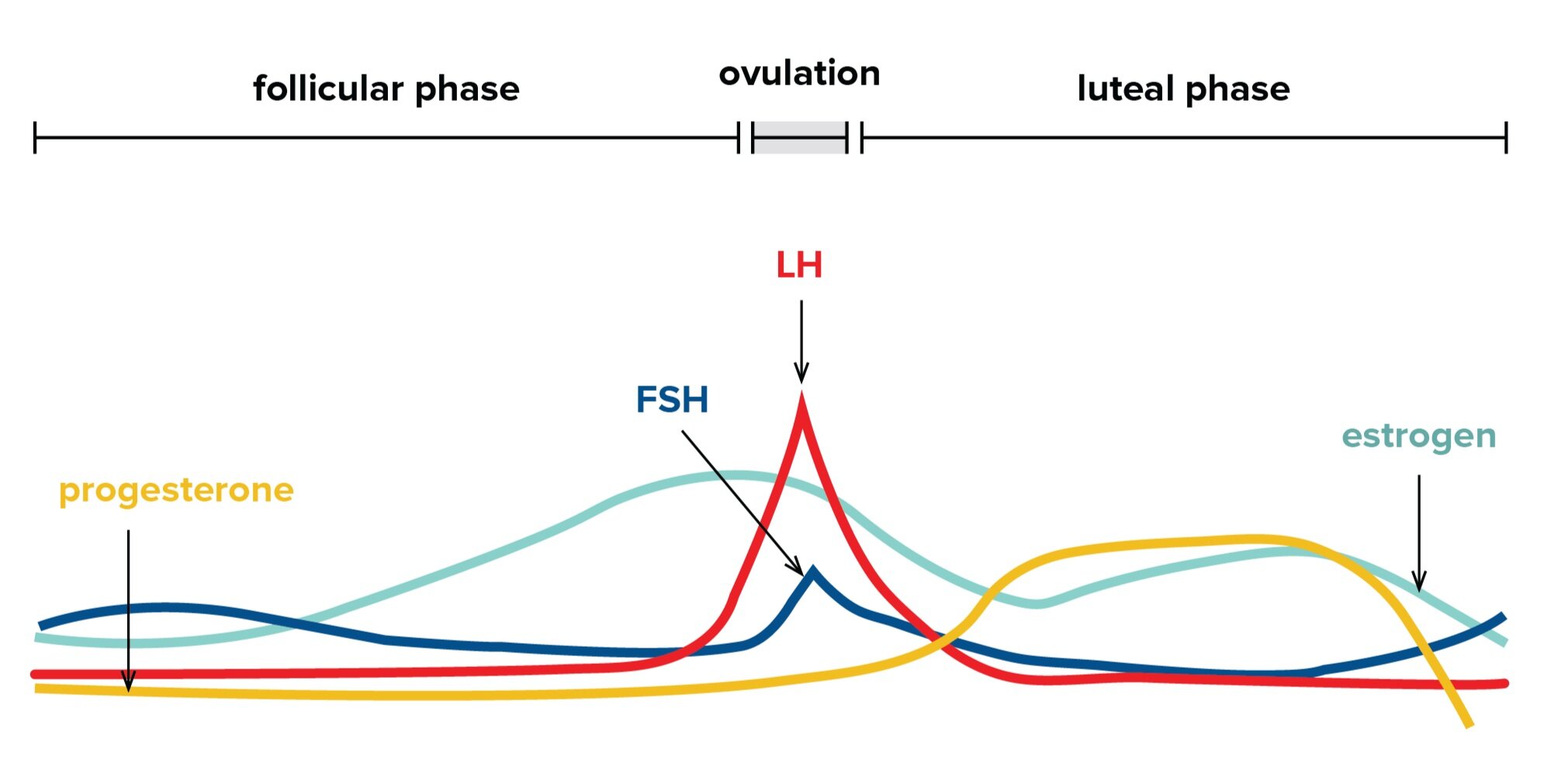
Figure 1 Changes in relative physiological concentrations of key endocrine hormones during the menstrual cycle.
In males, LH stimulates testosterone synthesis. During puberty, this hormone promotes the development of secondary sexual characteristics in males and stimulates sexual drive. However, this hormone is also crucial for a male fetus as it promotes the development of male genitalia.
In early development, early embryos are “sexually indifferent”: while their chromosomal sex is already determined, their genitalia and sex cells are not yet induced to become male or female. The presence of testosterone is crucial in inducing the early genitalia to follow a male course of development.
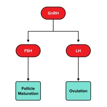
Figure 2 The HPG axis in females
Figure 3 The HPG axis in males
In normal physiology, male and female germ cells develop into different structures. Recall that the testes are male structures that store sperm, or male germ cells. The ovaries are female structures that store eggs, or female germ cells. These two sets of germ cells require specialized sets of helper cells to assist in their development: the development of sperm requires Sertoli cells and Leydig cells, while the development of oocytes requires granulosa cells and theca cells. The production of sperm and egg cells is collectively referred to as gametogenesis, as they result in the production of gametes.
b) Spermatogenesis
The male reproductive anatomy consists of both internal and external structures. The testes are the male gonads.
Spermatogenesis is the process through which sperm are formed in the seminiferous tubules of the testes. First, a diploid spermatogonium undergoes mitosis to form a diploid primary spermatocyte. After undergoing meiosis I, two haploid secondary spermatocytes are formed. Both of these then undergo meiosis II to form four haploid spermatids. The spermatids undergo further maturation and finally develop into spermatozoa.
This meiotic division and maturation occur in long, coiled structures within the testes called seminiferous tubules. Spermatogenesis is aided by Sertoli cells (specialized cells in the seminiferous tubules of the testes and are stimulated by FSH) and Leydig cells (cells that are also found in the testes and are stimulated by LH to produce testosterone).
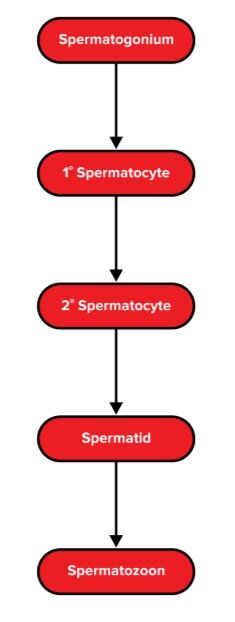
Figure 4 An overview of spermatogenesis
Sperm acquire motility and await ejaculation in a structure called the vas deferens. After maturation, each of these spermatids becomes a haploid spermatozoon. When ejaculation occurs, sperm will pass through the vas deferens, the ejaculatory duct, and then the urethra, which facilitates its exit from the penis.
Figure 5 The structure of the sperm cell.
At the head of the sperm cell, we find two important structures. First, we have the acrosome. Derived from the Golgi apparatus, it acts as a cap for the sperm and allows it to break the zona pellucida and penetrate the oocyte during fertilization. Second, we have the nucleus, which houses the DNA. Next, we have the midpiece portion of the sperm cell, which contains plenty of mitochondria. This allows the tail of the sperm, or flagellum, to have the energy it needs for locomotion. As the primary function of the sperm cell is to travel up the female reproductive tract and fertilize an oocyte, the sperm cell has evolved to have a high energy production capacity and locomotive ability.
c) Oogenesis
The female reproductive anatomy consists only of internal structures. The ovaries are the female gonads. In addition to producing estrogen and progesterone, the ovaries house ova, or eggs.
Oogenesis is the process through which oocytes are produced in females. While spermatogenesis begins around the onset of puberty in males, oogenesis begins before birth.
In utero, or before birth, diploid oogonia (ovarian stem cells) undergo mitosis to form diploid primary oocytes. These undergo meiosis and are stopped (or “arrested”) at prophase I until puberty. Thus, at birth, all of the oogonia a female has are primary oocytes that are arrested at prophase I.
During puberty, the meiotic division of these oocytes will continue. During each menstrual cycle, one primary oocyte is selected. This oocyte, which is encased in a follicle, will finish meiosis I and produce a haploid secondary oocyte and a much smaller polar body. These polar bodies contain very little cytoplasm and are discarded.
The secondary oocyte initiates meiosis II but stops at metaphase II. During ovulation, the follicle that the oocyte is encased in will rupture. The developing oocyte is released and eventually enters the Fallopian tubes.
It’s important to note that oocytes do not complete meiosis II until a sperm cell binds to the oocyte. Once a sperm cell binds, the oocyte will finish meiosis II and divide into two cells: a larger ovum which will undergo fertilization and a much smaller, second polar body.
Granulosa cells are found in the ovary and are stimulated by FSH to form the zona pellucida, a thick outer covering that protects the oocyte. Theca cells, which are also found in the ovaries, are stimulated by LH, and they produce androstenedione, a precursor for estrogen.
Figure 6 An overview of oogenesis.
Every month, one ovum is released into the abdominal cavity and enters one of two fallopian tubes, which serves as a link between the ovaries and the uterus. After a sperm cell is released into the cervix of the uterus during intercourse, it travels to the fallopian tube to fertilize the egg.
-----
Part 3: Fertilization and early development
a) Fertilization
Fertilization is the process through which a sperm cell and ovum fuse together. It typically occurs in the ampulla of the Fallopian tube, where it is widest. As we noted above, the sperm actually encounters a secondary oocyte. How is the meiotic division of the oocyte completed?
A set of reactions called the acrosomal reactions occur where acrosomal enzymes released by the cap on the sperm’s head allow it to penetrate the zona pellucida. The sperm then penetrates the cell membrane and releases its nucleus into the oocyte, allowing meiosis II to be completed. Now, the secondary oocyte has become an ovum.
A second reaction referred to as the cortical reaction occurs. The exact reaction is out of scope for the MCAT, but it increases calcium levels to prevent polyspermy, or the fertilization of an egg by more than one sperm. In humans, polyspermy is usually a lethal condition. Fertilization of an egg by two sperm results in a fused cell with ploidy 3N, which is nonviable for mitotic division (and thus, development).
After fertilization, we are left with one diploid cell with its own unique genetic material, a zygote. Recall that genetic sex is determined by the karyotype of the 23rd pair of chromosomes: where an XX karyotype leads to female development, and an XY karyotype leads to male development.
b) Cleavage, blastulation, and implantation
As the zygote begins to travel to the endometrium for implantation, it undergoes a series of cell divisions referred to as cleavage. This rapid cleavage is not exactly the same as normal mitotic division: while the genetic material is being duplicated and shared, the two resulting cells of each division do not appreciably grow. Our now multicellular organism, referred to as an embryo, will continue to divide until it forms a 16-cell ball called a morula. The morula is a solid sphere of packed cells with no way to differentiate the interior from the exterior.
The morula will drastically change and become a blastocyst through a process called blastulation. In contrast to the structurally homogeneous morula, the blastocyst has three main components. First is a hollow inner cavity that is filled with fluid, called the blastocoel. Second is the inner cell mass (ICM), an interior layer of cells that will ultimately develop into the fetus. Finally, the blastocyst contains trophoblast cells, which are found on the outside of the blastocyst.
The cells contained within the inner cell mass will develop into the many structures of the body. At this stage, these stem cells are considered to be pluripotent. This pluripotency is in contrast to the previously totipotent morula, which contains cells that can develop into either the body or the extraembryonic membranes. While totipotent cells can become any possible cell type, pluripotent stem cells can only become cells within the body.
As the process of blastulation occurs, the developing embryo travels down the Fallopian tube toward the uterus. When the blastocyst arrives at the uterus, the trophoblast cells penetrate the endometrium and form the placenta. The placenta is a fully functional endocrine organ that develops during pregnancy and is ejected after childbirth. While the process of placental development is out of scope for the MCAT, it’s important to recognize that the placental is a maternal and fetal organ that allows for the exchange of gases, nutrients, and antibodies—but does not allow for the mixing of blood.
c) Gastrulation
Once the blastula is implanted, gastrulation can soon occur. The process of gastrulation converts the ICM of the blastula into a gastrula and gives rise to the three germ cell layers. During this process, invagination occurs as some of the cells of the blastula begin to migrate into the blastocoel. This migration of cells, which occurs through a region called the primitive streak, is the first major movement of cells. This gives rise to the archenteron, which eventually becomes the gastrointestinal tract. (Recall that the gastrointestinal tract is essentially a long tube that runs through the body and is open throughout.) The archenteron’s opening, or blastopore, will become the anus in humans.
Figure 7 The development of zygote to gastrula.
The fate of the blastopore’s development depends on whether the organism is a deuterostome (meaning “second mouth”) or a protostome (“first mouth”). In deuterostomes, which include humans and other vertebrates, the blastopore gives rise to the anus. In protostomes, which include many invertebrates with bilateral symmetry, the blastopore will become the mouth.
-----
Part 4: Germ cell layers
The gastrula contains three distinct layers of cells—the endoderm, mesoderm, and ectoderm—arranged in a tubelike structure. This is in stark contrast to the structurally homogeneous ball of cells that formed the morula! Each of these three germ layers will ultimately develop into a different set of organs within the developed body.
a) Endoderm
The innermost germ layer, the endoderm, gives rise to what can be loosely defined as the interior of the body. The endoderm develops into the epithelial lining of the digestive and respiratory tracts, the liver, the pancreas, and the stomach. One important thing to note is that the epithelial lining of the digestive tract does not include the mouth or anus.
b) Mesoderm
Key areas of the mesoderm, or the middle layer of the gastrula, will give rise to specific systems and parts of the body. The mesoderm develops into somites, which later develop into muscles. Another portion develops into the lateral plate mesoderm, which later develops into the circulatory system, appendicular skeleton, the linings of body cavities. Other portions of the mesoderm develop into the gonads, kidneys, and adrenal cortex.
c) Ectoderm
At the outermost layer of the gastrula is the ectoderm. The ectoderm gives rise to what can be loosely defined as the exterior of the body—including the skin. This germ layer also develops into the nervous system and the epithelial lining of the mouth and anus.
Note that the nervous system develops from the ectoderm rather than a more “interior” germ cell layer. From an evolutionary standpoint, think of the brain and spinal cord as “modified skin.” The nervous system is formed through the invagination of the ectoderm in a process called neurulation, which we will discuss below.
Table 1 Summary table of the derivatives of the germ cell layers
| Germ cell layer | Derivatives |
|---|---|
Epithelial lining of respiratory tract Liver Pancreas Stomach |
|
Circulatory system Gonads Linings of body cavities Adrenal cortex |
|
Nervous system Epithelial lining of mouth and anus |
-----
Part 5: Neurulation
The process through which ectodermal cells give rise to the nervous system is known as neurulation. A structure known as the notochord first forms in the mesoderm. The notochord sends signals that induce the thickening of ectodermal cells, which then invaginate to form neural folds. These neural folds meet at the midline and close to form the neural tube. Later in development, the neural tube will develop into structures of the central nervous system: including the brain and spinal cord.
Figure 8 A cross-sectional view of neurulation.
It may be tempting to assume that the notochord will ultimately develop into the brain and spinal cord. In humans, this is not quite true—the notochord induces the development of these structures from the ectoderm while the notochord itself develops into the cartilaginous disks within the vertebral column.
When the neural folds fuse together to form the neural tube, certain adjacent cells migrate away and form the neural crest. These cells will move to the periphery and give rise to the structures, including the peripheral nervous system, teeth, cartilage, and specialized endocrine organs.
Figure 9 Formation of the neural tube and migration of neural crest cells.
By the end of neurulation, the developing embryo has a rudimentary nervous system and is beginning to develop organ systems. However, there is still a lot of growth and specialization of cells that need to happen before the embryo is a functional being!
As the embryo begins to grow into a fetus during the second and third trimesters of pregnancy, it will continue relying on maternal nutrients and the placenta for resources. These trimesters are periods of organ specialization, development, and growth.
-----
Part 6: Later development
a) Stages of pregnancy
Pregnancy, or gestation, is divided into three trimesters of roughly thirteen weeks each. During the first trimester, the embryo initiates organogenesis, or the development of organs. By the end of this trimester, most of the organs are developed; however, some organs, such as the lungs, continue developing even after birth.
In the second trimester, the embryo is now considered a fetus. During this phase of gestation, the fetus will undergo rapid growth as it develops a face, toes, and fingers.
In the third trimester, rapid growth and development continue. However, growth slows down as parturition, or childbirth, approaches. The beginning of this process is marked by the rupture of the amniotic sac: colloquially known as “water breaking.” This is then followed by uterine contractions to expel the fetus and placenta. Oxytocin, a peptide hormone, stimulates both uterine contractions during parturition and milk ejection during lactation.
For more information on the function of these hormones, be sure to refer to our guide on the endocrine system.
b) Cell specialization
Cell specialization is the process by which a cell changes into a specific type of cell. The first step of this process is determination, where a cell irreversibly commits to a certain cell type. This step precedes differentiation, which occurs as a cell undergoes structural and biochemical changes to carry out the functions of its cell type.
Cell specialization can occur via multiple mechanisms. For example, an uneven distribution, or gradient, of mRNA during mitosis may drive differentiation as it can lead to selective transcription. Additionally, cell-cell communication is another common way by which cells differentiate. A cell can release morphogens, which are signaling molecules that promote differentiation. Any cell releasing the morphogens is known as an inducer, while the receiving cell is a responder.
After specialization, each cell must be in its correct spatial location. During development, the organized migration of cells to specific anatomical locations is crucial. The previous discussion on the movement of neural crest cells during neurulation is a classic example of developmental cell migration. Errors during this process can give rise to various calamities, such as intellectual disabilities.
c) Apoptosis and regeneration
Apoptosis refers to programmed cell death in multicellular organisms. First, a cell breaks down to form apoptotic blebs, which are membrane-bound sacs containing cellular material. These are then broken down into apoptotic bodies, which are digested by other cells.
Apoptosis is crucial for development as the targeted death of specific cells allows the fetus to form into a specific shape. For instance: your fingers, which are now separated, were once webbed together. During fetal development, however, the cells forming the webbing performed apoptosis, thus dying and allowing for separation between the figures.
In addition to controlled cell death, humans also possess regenerative abilities. The liver, for example, is an organ that can regenerate even when up to 60% is removed. Thus, a living person can donate part of their liver and have it regenerate.
Relative to other species, however, regeneration is quite limited in humans as we are capable of only incomplete regeneration. Organisms capable of complete regeneration store stem cells, which can then be used to regrow entire body parts when necessary.
-----
Part 7: High-yield terms
HPG axis: axis of endocrine control consisting of the hypothalamus, anterior pituitary gland, and gonads
GnRH: gonadotropin-releasing hormone; endocrine hormone released by the hypothalamus
FSH: follicle-stimulating hormone; endocrine hormone released by the anterior pituitary gland
LH: luteinizing hormone; endocrine hormone released by the anterior pituitary gland
Follicle: immature cells that assist the development of oocytes in females
Spermatogenesis: the process through which sperm are formed in the seminiferous tubules of the testes
Sertoli cells: specialized cells in the seminiferous tubules of the testes and are stimulated by FSH
Leydig cells: cells found in the testes and are stimulated by LH to produce testosterone
Oogenesis: the process through which oocytes are produced in females
Polar body: a cell resulting from meiotic division of the oocyte; contains very little cytoplasm and is discarded
Granulosa cells: cells found in the ovary and are stimulated by FSH to form the zona pellucida
Theca cells: cells found in the ovaries; stimulated by LH and produce estrogen
Acrosomal reactions: reactions in which acrosomal enzymes released by the cap on the sperm’s head allow it to penetrate the zona pellucida
Cortical reaction: reactions that increase calcium levels in order to prevent polyspermy
Morula: structurally homogeneous ball of packed cells
Blastocyst: contains hollow blastocoel, inner cell mass, and trophoblast cells
Gastrula: inner cell mass that has differentiated into the endoderm, mesoderm, and ectoderm
Archenteron: invagination of the gastrula that develops into gastrointestinal tract
Neurulation: process through which ectodermal cells give rise to the nervous system
-----
Part 8: Passage-based questions and answers
Egg activation refers to events required for the transformation of gametes into an embryo, including the establishment of the polyspermy block, completion of meiosis, entry into mitosis, selective recruitment and degradation of maternal mRNA, and pronuclear development.
Researchers wish to determine whether fluxes in zinc concentration occur during human egg activation. Researchers first treated human eggs with the calcium transporter, Ca-ionomycin, which delivers a bolus of exogenous calcium directly into the egg and bypasses the sperm-induced signaling cascades needed to elicit a rise in endogenous stores of intracellular calcium. Intracellular calcium and extracellular zinc dynamics in individual eggs were monitored by live-cell imaging using fluorescent microscopy. The results were quantified and are shown in Figure 1.
Figure 1
Question 1: What is the ploidy of a mature unfertilized female gamete?
A) 4n
B) 3n
C) 2n
D) n
Question 2: Which of the following most accurately describes the results presented in Figure 1?
A) Egg activation is associated with a rise in both zinc and calcium levels
B) Egg activation is associated with a decrease in both zinc and calcium levels
C) Egg activation is associated with a decrease in zinc levels but an increase in calcium levels
D) Egg activation is associated with an increase in zinc levels but a decrease in calcium levels
Question 3: Which of the following processes assist in preventing polyspermy?
A) Fertilization
B) Acrosomal reactions
C) Cortical reactions
D) Mitosis
Question 4: Why did the researchers use Ca-ionomycin?
A) It fertilizes the egg, thereby inducing a rise in intracellular calcium
B) It bypasses the need for sperm to induce the intracellular rise in calcium levels
C) It is a zinc analog that can trigger egg activation
D) It is a calcium analog that can trigger egg activation
Question 5: Which of the following is not true of gamete production in females?
A) A diploid cell ultimately results in the formation of a haploid cell
B) The process begins at the onset of puberty
C) The process involves both mitosis and meiosis
D) The process also produces polar bodies
Answer key for passage-based questions
Answer choice D is correct. A mature unfertilized female gamete is a haploid cell (choice D is correct). This means it has a ploidy of 1n. A primary oocyte undergoes the first meiotic division and is ploidy 2N, while the secondary oocyte has ploidy of 1N. The secondary oocyte is ultimately fertilized and completes the second meiotic division after fertilization.
Answer choice A is correct. According to Figure 1, there is a directly proportional relationship between zinc and calcium levels (choices C and D are incorrect). Further, the passage states that calcium is associated with egg activation. Thus, egg activation is associated with an increase in zinc and calcium levels (choice B is incorrect).
Answer choice C is correct. The cortical reactions help prevent polyspermy. Fertilization is the joining of a sperm cell and egg cell (choice A is incorrect). The acrosomal reactions allow the sperm cell to penetrate the zona pellucida (choice B is incorrect). Mitosis is a cellular process through which cells divide to form identical daughter cells (choice D is incorrect).
Answer choice B is correct. The passage states that the Ca-ionomycin is a calcium transporter (choices C and D are incorrect). The passage also states that it bypasses the sperm-induced signaling cascades needed to increase calcium levels (choice A is incorrect). Under normal conditions, large amounts of intracellular calcium must be released to complete fertilization.
Answer choice B is correct. In females, oogenesis begins in utero with the formation of primary oocytes (choice B is correct). The meiotic division of oogonia results in the formation of diploid primary oocytes, which undergo meiotic division to form haploid germ cells (choice A is incorrect). Both mitosis and meiosis are necessary processes in the formation of mature ova (choice C is incorrect). The first and secondary meiotic divisions result in the formation of polar bodies, which are nonfunctional sister cells (choice D is incorrect).
-----
Part 9: Standalone questions and answers
Question 1: Which of the following make up the HPG axis?
I. Gonadotropin-releasing hormone
II. Luteinizing hormone
III. Growth hormone-releasing hormone
A) I only
B) II only
C) I and II
D) I, II, and III
Question 2: What is the function of trophoblast cells?
A) To develop into the placenta
B) To become the developing nervous system
C) To support theca cells
D) None of the above
Question 3: Which of the following does NOT originate from the endoderm?
A) The liver
B) The epithelial lining of the digestive tract
C) The epithelial lining of the respiratory tract
D) The epithelial lining of the mouth
Question 4: Which of the following structures develops into the peripheral nervous system?
A) Notochord
B) Neural folds
C) Neural crest cells
D) Neural tube
Question 5: What is the purpose of the acrosomal reactions?
A) To protect sperm cells from an acidic environment
B) To generate energy for sperm cells
C) To allow sperm cells to penetrate the zona pellucida
D) To prevent polyspermy
Question 6: A male patient at a fertility clinic is noted to have an unusually low sperm count. What types of cells should be suspected to be dysfunctional?
A) Leydig cells
B) Sertoli cells
C) Granulosa cells
D) Theca cells
Answer key for standalone questions
Answer choice C is correct. The HPG axis begins at the hypothalamus with the release of GnRH (choice I is correct). GnRH acts on the anterior pituitary gland to release FSH and LH, which in turn act on the gonads (choice II is correct). Growth hormone-releasing hormone is not a part of the HPG axis (choice III is incorrect).
Answer choice A is correct. Trophoblast cells will penetrate the endometrium and eventually give rise to the placenta (choice A is correct). The inner cell mass will become the fetus (choice B is incorrect). Theca cells are a type of support cell that nourish developing oocytes (choice C is incorrect).
Answer D is correct. The endoderm gives rise to the epithelial lining of the digestive and respiratory tract, as well as its associated viscera (choices A, B, and C are incorrect). The endoderm does not give rise to the epithelial lining of the mouth or anus, which are derived from the ectoderm (choice D is correct).
Answer choice C is correct. The notochord is a structure within the mesoderm that induces change within the overlying ectoderm (choice A is correct). Signals from the notochord induce the formation of neural folds, which come together to form the neural tube. The neural tube is a precursor to the central nervous system (choices B and D are incorrect). Neural crest cells migrate away from the neural tube and ultimately develop into the peripheral nervous system, teeth, and certain endocrine organs (choice C is correct).
Answer choice C is correct. The acrosomal reactions are a set of enzymatic reactions that allow a sperm cell to digest and penetrate the zona pellucida of the egg cell (choice C is correct). The acrosomal reactions do not serve to protect a sperm cell from an acidic environment (choice A is incorrect). The mitochondria found in the midpiece are responsible for generating energy for the sperm cell (choice B is incorrect). The cortical reaction prevents polyspermy, or the fertilization of an egg by multiple sperm (choice D is incorrect).
Answer choice B is correct. Sertoli cells are support cells that assist in the synthesis of sperm and should be suspected to be dysfunctional if the patient is unable to produce motile sperm (choice B is correct). Leydig cells produce testosterone (choice A is incorrect). Granulosa cells form the zona pellucida (choice C is incorrect). Theca cells produce androstenedione, an estrogen precursor (choice D is incorrect).