
Endocrine System for the MCAT: Everything You Need to Know
A complete guide to the endocrine system on the MCAT, covering key hormones, physiological effects, and practice questions to boost your biology score.

(Note: This guide is part of our MCAT Biology series.)
Part 1: Introduction to the endocrine system
Part 2: Classifying hormones
a) Peptide hormones
b) Steroid hormones
c) Tyrosine derivatives
Part 3: Endocrine Glands and Secreted Hormones
a) Pancreas
b) Anterior and posterior pituitary glands
c) Thyroid and parathyroid glands
d) Adrenal cortex and adrenal medulla
Part 4: Hormonal Axes
a) Hypothalamic-pituitary-adrenal (HPA) axis
b) Hypothalamic-pituitary-gonadal (HPG) axis
Part 5: High-Yield Terms
Part 6: Passage-Based Questions and Answers
Part 7: Standalone Questions and Answers
----
Part 1: Introduction to the endocrine system
The endocrine system works with the nervous system to alter metabolism, guide reproduction, and regulate growth. In this guide, we will provide you with an overview of endocrine-based content you need to know for the MCAT. At the end of this guide, there is an MCAT-style practice passage and standalone questions to test your knowledge and show you how the AAMC likes to ask questions.
----
Part 2: Classifying hormones
When functioning correctly, the endocrine system maintains homeostasis in the body. In other words, hormones are released in response to a condition rather than to create one. For example, insulin is released in response to high blood sugar levels; it is not released to create low blood sugar. This mechanism of action is referred to as negative feedback. While positive feedback is a mechanism to stimulate a condition through an action, negative feedback seeks to lessen a condition through an action.
How does the endocrine system accomplish this? The endocrine system releases hormones into the body’s general circulation, most often through blood, to affect other cells or tissues at a distance. (This is in contrast to autocrine signaling, in which a signaling molecule is released to affect the same cell or tissue, and paracrine signaling, in which a signaling molecule is released to affect a neighboring cell or tissue.)
Hormones can be classified into three distinct types, based on their structure: peptide hormones, steroid hormones, and tyrosine derivatives.
a) Peptide hormones
Peptide hormones are derived from peptides: short amino acid chains linked together by peptide bonds (e.g., O=C-NH bonds). Peptides are essentially short proteins. Therefore, peptide hormones are generally polar and water-soluble, allowing them to move freely through the bloodstream. However, peptides cannot easily diffuse through the nonpolar cell membrane. Therefore, peptide hormones generally require the presence of a receptor on the surface of the cell in order to effect an action.
When the peptide hormone binds to the receptor, there are multiple ways the receptor may respond. The receptor may act as an ion channel or interact with other proteins to create an ion-channel effect. (You can find more information on this signaling in our guide on lipids and cell membranes.)
The MCAT will most likely test your knowledge of a specific receptor system known as a G-protein coupled receptor, or GPCR. Upon binding to the hormone, G protein-coupled receptors undergo a conformational change that releases a G-protein by exchanging a low-energy GDP for a high-energy GTP. The G-protein activates the secondary messenger “cascade,” allowing secondary messengers like cAMP or IP3 to initiate a “cascade” of reactions within the cell. These secondary messengers serve to greatly amplify the effect of the hormone, as the binding of a single hormone molecule on the surface of the cell can result in a much higher concentration of secondary messengers to be activated!

Figure 1 An overview of GPCR function.
Here is a list of the specific peptide hormones you’ll need to study for the MCAT. We’ll cover their functions later in this guide.
Table 1 Key peptide hormones and their production location
| Anterior pituitary | Posterior pituitary | Parathyroid | Pancreas | Thyroid |
|---|---|---|---|---|
| FSH LH ACTH TSH Prolactin Endorphins Growth Hormones |
ADH Oxytocin |
PTH | Glucagon Insulin |
Calcitonin |
b) Steroid hormones
Steroid hormones are derived from cholesterol and are considered lipids. Recall that lipids are nonpolar and so are not soluble in water or blood. In the bloodstream, steroid hormones require a protein transport molecule to carry them.
In contrast to peptide hormones, they are able to directly diffuse through cell membranes. As a result, they do not require the presence of a receptor system on the cell membrane to effect their action. Steroid hormones directly pass through the membrane and can bind to receptors in the cytosol, be transported to the nucleus, and affect transcription by binding to DNA.
Organized by their glandular origin, here are the steroid hormones you’ll need to study for the MCAT.
Table 2 Key steroid hormones and their production location
| Adrenal Cortex | Gonads (Testes and Ovaries) |
|---|---|
Aldosterone *More detailed coverage in our guide on the digestive and excretory systems |
Testosterone Progesterone *More detailed coverage in our guide on reproduction and development |
c) Tyrosine derivatives
Tyrosine derivative hormones are also lipid soluble and require a protein transport molecule to be carried in the blood. As you might guess, they are modified versions of the amino acid tyrosine. This means that they are very small hormones in contrast to peptide and steroid hormones.
Organized by their glandular origin, here are the tyrosine derivative hormones you’ll need to study for the MCAT.
Table 3 Tyrosine derived hormones and their production location
| Thyroid | Adrenal Medulla |
|---|---|
T4 (Contains 4 iodine atoms) |
Norepinephrine (Both categorized as catecholamines) |
----
Part 3: Endocrine glands and secreted hormones
There is an important distinction to be made between the endocrine and exocrine systems. Exocrine systems release enzymes into external environments through ducts; sweat, mucous, oils are common examples of exocrine glands. Meanwhile, endocrine systems release hormones into the bloodstream. Because hormones are released into the blood (e.g., general circulation), they must first travel and bind to highly specific receptors to have an effect. Therefore, the impact of endocrine glands impacts is slower-moving and longer-lasting.
The endocrine system is also intimately connected with the nervous system. Recall that the endocrine system arises from neural crest cells, which give rise to the peripheral nervous system and many other associated structures. (For more information on this, be sure to refer to our guide on reproduction and development.)
As you work through this section of the guide, be sure to keep this relationship between the nervous and endocrine systems in mind. Several hormones may also function as neurotransmitters, and secretions from endocrine glands may have negative or positive feedback effects on the central nervous system.
a) Pancreas
The pancreas is an organ that acts both as an exocrine and an endocrine gland. For its exocrine functions, it secretes digestive enzymes like trypsin and amylase through the pancreatic duct. For its endocrine functions, the pancreas releases insulin and glucagon into our bloodstream to regulate blood sugar levels.
The pancreas contains specialized clusters of cells called the Islets of Langerhans. Within these “islets,” there are three important cell types to know. Each of the cell types—β-cells, α-cells, and δ-cells—secretes a different hormone with a different effect.
Insulin is a peptide hormone that is released by β-cells of the pancreas in response to high levels of carbohydrates and protein in the blood. In response to insulin, carbohydrates are stored in the liver and muscles as glycogen and fat are stored in adipose tissue. Therefore, insulin lowers blood glucose.
Glucagon and insulin are opposites. Glucagon is a peptide hormone that is released by α-cells of the pancreas in response to low nutrient levels in the blood. Its primary function is to stimulate glycogenolysis (the breakdown of glycogen) and gluconeogenesis (the generation of glucose) in the liver to raise blood glucose levels.
Somatostatin is a peptide hormone that is released by δ-cells of the pancreas. Somatostatin is a hormone that blocks the action of other hormones—including insulin, glucagon, and growth hormones. Its primary function is to slow the activity of other endocrine signaling and lower the metabolic rate of the body.
You can find more information on the actions of insulin and glucagon in our guide on carbohydrate metabolism.
b) Anterior and posterior pituitary glands
The pituitary gland is a small structure nestled within our brain. It is divided into two smaller structures: the anterior and posterior pituitary glands, both of which have unique functions. The hypothalamus, considered to be the master control gland of the body, regulates the anterior and posterior pituitary in different ways.
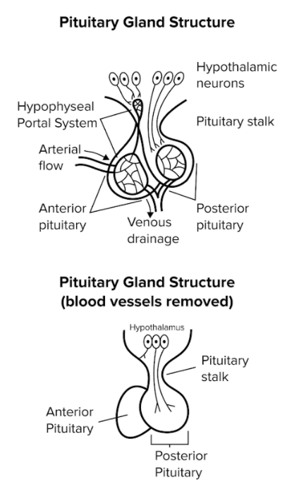
Figure 2 The hypothalamus interacts with the anterior pituitary gland through a portal system.
The hypothalamus interacts with the posterior pituitary through neurons. These nerve cells synthesize oxytocin and ADH (antidiuretic hormone, also known as vasopressin). Oxytocin and ADH travel down neuron axons from the hypothalamus, through the pituitary stalk, and into the posterior pituitary, where they are released into the blood.
The hypothalamus interacts with the anterior pituitary gland through a portal system, or network of intertwined blood vessels and capillaries. This particular portal system is known as the hypophyseal portal system. The hypothalamus secretes its own set of hormones into the hypophyseal portal system to signal the anterior pituitary to take action.
These hormones that are secreted by the hypothalamus bind to receptors in the anterior pituitary to stimulate the release of other hormones. Recall that the anterior pituitary gland secretes the “FLAT PEG” hormones: FSH, LH, ACTH, TSH, prolactin, endorphins, and growth hormone. Below, we’ll take a short tour of each of these hormones and their roles.
FSH stands for “follicle-stimulating hormone.” In women, FSH stimulates the maturation of follicles into mature eggs; in men, FSH triggers the maturation of sperm. LH stands for “luteinizing hormone.” It stimulates the production of androgens and estrogens, including testosterone. In women, LH also stimulates progesterone production. Both FSH and LH play important roles in the menstrual cycle. More information on this can be found in our guide on reproduction and development.
Adrenocorticotropic hormone (ACTH) is released in response to stress—either metabolic stress or stress from the environment. ACTH stimulates the adrenal cortex to release cortisol, a type of stress hormone. (You may also see cortisol referred to as a glucocorticoid.) As a result, cortisol is produced by the body in response to stress.
Prolactin stimulates the production of milk, or lactation, from mammary glands. It should not be confused with oxytocin, which leads to the release of milk. Normally, progesterone and estrogen inhibit milk production, but suckling stimulates the hypothalamus, which in turn stimulates the anterior pituitary to release prolactin.
Endorphins are natural painkillers released in response to pain or physical stress, for instance, after you stub your toe. Endorphins can also be released in response to exercise, leading to a so-called “runner’s high.”
True to its name, human growth hormone (hGH) stimulates growth and development, including the increase of cell size, muscle size, and rate of protein synthesis. Its release is stimulated by growth hormone-releasing hormone (GHRH) from the hypothalamus. (There’s an important distinction between human growth hormone and human chorionic growth hormone (hCG), which is produced by an early embryo during pregnancy.)
The posterior pituitary gland secretes oxytocin and ADH. Oxytocin leads to the release of milk after it has been produced in the mammary glands and also increases the frequency of uterine contractions during childbirth. Antidiuretic hormone, also known as ADH or vasopressin, increases the permeability of the collecting ducts of the kidney to water. It allows more water to be resorbed, concentrating urine while decreasing urine volume. Another important effect of ADH is that it increases blood pressure, as more fluid is resorbed into the bloodstream.
c) Thyroid and parathyroid glands
Although T3 has 3 iodine atoms and T4 has 4 iodine atoms, T3 and T4 work in tandem to accomplish the same function. They increase the body’s basal (resting) metabolic rate. T3 and T4 are lipid soluble and are regulated by TSH (a hormone released by the anterior pituitary).
Calcitonin stimulates osteoblasts to build bone, which decreases blood calcium. Its action is opposite to parathyroid hormone (PTH), a hormone secreted by the parathyroid glands. Parathyroid hormone (PTH) stimulates osteoclasts to consume bone, which increases blood calcium. More information on calcitonin and PTH can be found in our guide on the musculoskeletal system.
d) Adrenal cortex and adrenal medulla
The adrenal glands are found sitting atop the kidneys. The adrenal cortex refers to the outside layer of the gland, while the adrenal medulla refers to its internal structure. The adrenal cortex secretes aldosterone and cortisol. The adrenal medulla secretes epinephrine and norepinephrine: both tyrosine derivatives known as catecholamines.
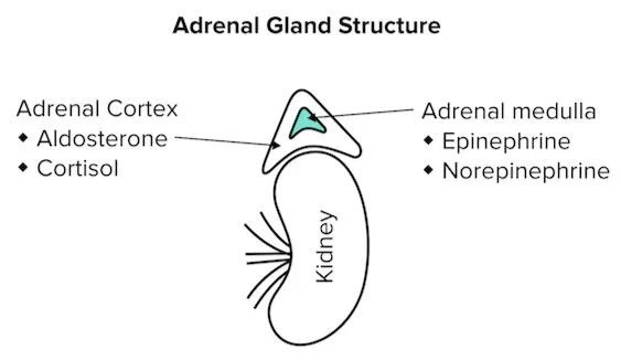
Figure 3 The structure of the adrenal gland has 2 main components: the cortex and medulla
Cortisol belongs to a subclass of steroid hormones known as glucocorticoids. Glucocorticoids increase blood glucose levels and the rate of protein/fat metabolism. In particular, cortisol is a stress hormone that stimulates gluconeogenesis in the liver to increase blood glucose levels. Cortisol also has anti-inflammatory properties that reduces the immune system response. These effects have evolved to mitigate the effects of stress when the body needs to produce energy and prevent inflammation that could slow us down.
Epinephrine and norepinephrine (also called adrenaline and noradrenaline) are tyrosine derivatives categorized as catecholamines and are synthesized in the adrenal medulla. Epinephrine is considered a hormone, while norepinephrine is a neurotransmitter. Both are released in response to stress. When faced with stress, the sympathetic nervous system stimulates the secretion of epinephrine and norepinephrine. This, in turn, stimulates vasoconstriction of the gastrointestinal tract and kidneys, along with the vasodilation of lungs, brain, heart, and skeletal muscle. Together, these physiological effects create the “fight-or-flight” response.
----
Part 4: Hormonal axes
The MCAT covers 3 hormonal axes:
1. The hypothalamic-pituitary-adrenal (HPA) axis
2. The hypothalamic-pituitary-gonadotropin (HPG) axis
3. The renin-angiotensin-aldosterone system (RAAS)
This guide will cover the hypothalamic-pituitary-adrenal axis (HPA) axis and the hypothalamic-pituitary-gonadal (HPG) axis. You can find more information on the renin-angiotensin-aldosterone system in our guide on the digestive and excretory systems.
a) Hypothalamic-pituitary-adrenal (HPA) axis
The HPA axis is our body’s primary stress response system. In response to environmental stressors, the hypothalamus releases corticotropin-releasing hormone (CRH). CRH is a peptide hormone that stimulates ACTH production in the anterior pituitary. Then, ACTH stimulates the adrenal cortex to release cortisol, which ultimately stimulates gluconeogenesis and the breakdown of proteins while decreasing inflammation.
The HPA axis is regulated via a negative feedback loop, where the presence of cortisol inhibits CRH secretion by the hypothalamus and ACTH secretion by the anterior pituitary.
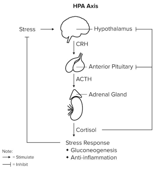
Figure 4 Feedback loops of the HPA axis.
b) Hypothalamic-pituitary-gonadal (HPG) axis
The HPG axis plays an important role in the reproductive system. Once again, the axis starts at the hypothalamus, which secretes gonadotropin-releasing hormone (GnRH). GnRH stimulates the production of FSH and LH in the anterior pituitary, which in turn act on the gonads to produce more testosterone (in males) or estrogen/progesterone (in females).
Similar to the HPA axis, the HPG axis is also regulated via a negative feedback loop. The presence of testosterone, estrogen, or progesterone in the bloodstream inhibits GnRH secretion in the hypothalamus.
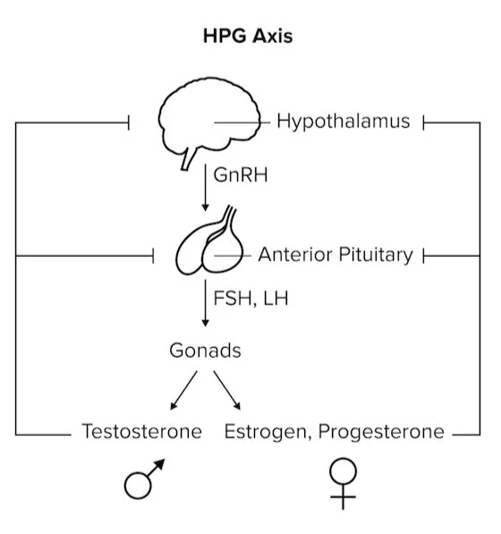
Figure 5 Feedback loops of the HPG axis.
----
Part 5: High-yield terms
GPCR: G-protein coupled receptor
FSH: follicle-stimulating hormone
LH: luteinizing hormone
ACTH: adrenocorticotropic hormone
TSH: thyroid-stimulating hormone
GHRH: growth hormone-releasing hormone
hGH: human growth hormone
hCG: human chorionic growth hormone
ADH: antidiuretic hormone, or vasopressin
T4: thyroxine
PTH: parathyroid hormone
Catecholamines: tyrosine derivatives that include epinephrine and norepinephrine
HPA: hypothalamic-pituitary-adrenal axis
HPG: hypothalamic-pituitary-gonadal axis
RAAS: renin-angiotensin-aldosterone system
CRH or CRF: corticotropin-releasing hormone/factor
----
Part 6: Passage-based questions and answers
The pituitary gland works closely with the hypothalamus to regulate homeostasis, metabolism, reproduction, and growth in multiple target organs. The pituitary gland is separated into the adenohypophysis and neurohypophysis, known as the anterior and posterior lobes, respectively. The anterior pituitary secretes five distinct types of hormones: somatotropes (hGH), lactotropes (PRL, or prolactin), thyrotropes (TSH), corticotropes (ACTH), and gonadotropes (FSH, LH).
Pituitary organogenesis and hormone synthesis is a complex process that is not completely understood. Many different transcription factors are temporally and spatially regulated. Pit-1, for example, is required for pituitary cell differentiation, and the Pit1 gene has been implicated in GH, TSH, and prolactin deficiency.
In a recent study, scientists have discovered a transcription factor, ZBTB20, that could play a key role in anterior pituitary development and lactotrope specification in mice. ZBTB20 is a zinc finger protein that contains BTB/POZ domains and C2H2 Kruppel-type zinc fingers.
To further characterize the role of ZBTB20 in anterior pituitary development, scientists performed a knockout experiment in mice. ZBTB20 global knockout mice were bred and their phenotypes characterized. Hypopituitarism was found to be a result of the ZBTB20 knockout. Further results of this study are presented in Figure 1.
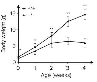
Figure 1
Researchers followed the mice throughout their lifespan. At postnatal day 21, or P21, scientists performed ELISA experiments to analyze anterior pituitary hormonal contents in both wildtype and knockout mice. The results are presented in figure 2.
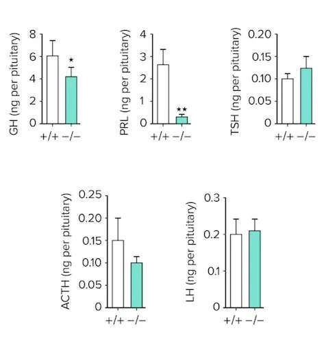
Figure 2 GH: growth hormone; PRL: prolactin; TSH: thyroid-stimulating hormone; ACTH: adrenocorticotropic hormone; LH: luteinizing hormone
Question 1: Suppose a patient presents unregulated over-secretion of thyrotropes from the anterior pituitary. Which of the following symptoms is this patient most likely to experience?
A) Weight loss
B) Vasodilation of heart and lungs
C) Increased sex hormone levels
D) Gigantism
Question 2: Abnormal levels of which hormone most likely explain the result shown in Figure 1?
A) ACTH
B) Prolactin
C) TSH
D) hGH
Question 3: Which of the following hormones are secreted by the neurohypophysis?
A) Epinephrine
B) Oxytocin
C) Progesterone
D) Aldosterone
Question 4: Which of the following conclusions can be drawn from Figure 2?
A) ZBTB20 knockout leads to decreased production in all hormones from the anterior pituitary.
B) ZBTB20 knockout has little to no effect on mice.
C) ZBTB20 knockout leads to severe defect of lactotropes.
D) ZBTB20 is necessary for anterior pituitary organogenesis.
Answer key for passage-based questions
2. Answer choice D is correct. The passage states that a knockout of ZBTB20 leads to hypopituitarism. Based on the figure, the body weight of ZBTB20 knockout mice is significantly lower than the control mice. Of the hormones presented, only low levels of growth hormone can contribute to this result (choice D is correct). Deficiencies in adrenocorticotropic hormone (ACTH), prolactin, or thyroid-stimulating hormone would be unlikely to produce these results.
3. Answer choice B is correct. The neurohypophysis refers to the posterior pituitary. The posterior pituitary only secretes ADH and oxytocin (choice B is correct). Epinephrine is secreted by the adrenal medulla (choice A is incorrect). Progesterone is secreted by the gonads (choice C is incorrect). Aldosterone is secreted by the adrenal cortex (choice D is incorrect).
4. Answer choice C is correct. Based on the information presented in Figure 2, it is clear that there is almost no prolactin present in ZBTB20 knockout mice compared to wild-type mice (choice C is correct). There is also a statistically significant decrease in growth hormone, but all other hormone levels appear to be similar. The lack of asterisk above the bars shows they are not different in a statistically significant way (choices A and B are incorrect). An anterior pituitary is still formed in ZBTB20 knockouts, as evidenced by hormone production (choice D is incorrect).
----
Part 7: Standalone questions and answers
Question 1: Suppose a patient receives a large dose of vasopressin. Which of the following effects are most likely to take place?
I. Increase in blood pressure
III. Increase in urine osmolarity
A) I only
B) I and III
C) II and III
D) I, II, and III
Question 2: Which of the following responses is LEAST likely in response to stimulation of the sympathetic nervous system?
A) Release of catecholamines from the adrenal medulla
B) Release of corticotropin-releasing hormone (CRH) from the hypothalamus
C) Vasoconstriction of the lungs and skeletal muscle
D) Increase of gluconeogenesis in the liver
Question 3: Researchers develop a drug that inhibits the exocrine function of the pancreas. The primary action of this drug will most likely:
A) Decrease the amount of trypsin released into the blood
B) Decrease the amount of amylase released through a duct
C) Decrease the amount of insulin released into the blood
D) Decrease the amount of glucagon released through a duct
Question 4: Which of the following hormones does the anterior pituitary gland NOT produce?
A) Human growth hormone
B) Luteinizing hormone
C) Thyroid-stimulating hormone
D) Antidiuretic hormone
Question 5: Which of the following statements about peptide hormones is FALSE?
A) Peptide hormones usually directly affect transcription
B) Peptide hormones are water-soluble
C) Peptide hormones can act through secondary messengers
D) Peptide hormones are derived from proteins
Answer key for standalone questions
2. Answer choice C is correct. Stimulation of the sympathetic nervous system leads to activation of the “fight-or-flight” response. In response to stress, the adrenal medulla synthesizes epinephrine and norepinephrine, or the catecholamines (choice A is incorrect). These catecholamines work to vasodilate the lungs and skeletal muscle (choice C is correct). Another response to stress is produced by the hypothalamic-pituitary-adrenal (HPA) axis. The hypothalamus releases cortico-tropin releasing hormone (CRH), increasing ACTH, which increases cortisol (choice B is incorrect). Cortisol increases gluconeogenesis in the liver (choice D is incorrect).
3. Answer choice B is correct. Keep in mind the difference between exocrine and endocrine functions. Exocrine function leads to the release of enzymes into external environments, while endocrine function leads to the release of hormones into the blood (choices A and C are incorrect). Trypsin and amylase are digestive enzymes secreted by the pancreas (choice B is correct), while insulin and glucagon are endocrine hormones released by the pancreas into the bloodstream (choice D is incorrect).
4. Answer choice D is correct. Recall the “FLAT PEG” hormones: the anterior pituitary gland secretes FSH, LH, ACTH, TSH, prolactin, endorphins, and growth hormone. Antidiuretic hormone (ADH) and oxytocin are both secreted by the posterior pituitary (choice D is correct).
5. Answer choice A is correct. Steroid hormones usually act at the transcription level, as they can diffuse through cell membranes to bind to receptors in the cytosol and be transported to the nucleus (choice A is correct). Peptide hormones are derived from proteins (choice D is incorrect) and are therefore water-soluble, which means they can travel freely in the blood but cannot diffuse through cell membranes (choice B is incorrect). Therefore, the effects of peptide hormones are receptor-mediated, which means they can act through a secondary messenger like cAMP, cGMP, or calmodulin (choice C is incorrect).