
Immune System for the MCAT: Everything You Need to Know
Master MCAT immune system concepts with high-yield content, targeted practice questions, and detailed answers to boost your understanding and score.
(Note: This guide is part of our MCAT Biology series.)
Table of Contents
Part 1: Introduction
Part 2: Innate vs adaptive immunity
Part 3: Innate Immune System
a) Anatomical barriers
b) Cellular component
c) Complement system
d) Cytokines
e) Inflammation
Part 4: Adaptive Immune System
a) B cells and humoral immunity
b) T cells and cytotoxic immunity
Part 5: Anatomy of the Immune System
a) Bone marrow
b) Spleen
c) Thymus
d) Lymphatic system
Part 6: Antigens and Antibodies
a) Antibody structure
b) Antigen presentation
c) Recognition of self and non-self
Part 7: High-Yield Terms
Part 8: Passage-Based Questions and Answers
Part 9: Standalone Questions and Answers
-----
Part 1: Introduction
The immune system is responsible for protecting the body against foreign invaders and diseases. It is a complex system that relies on constant feedback to develop immunity. There are 2 major components of the immune system that we will discuss: innate and adaptive immunity. We will also learn about other components of the body that aid in the immune response, as well as how immune system components eliminate diseases. Overall, this is a low to medium yield topic, but various aspects are regularly tested in passages and questions.
-----
Part 2: Innate vs adaptive immunity
The highest level of classification within the immune system is between the innate immune response and the adaptive immune response. These will be discussed in greater detail in the next two parts. In general, the innate immune response is composed of bodily components that non-selectively target diseases and foreign pathogens, while the adaptive immune response is selective to certain diseases. You are born with your innate immune response ready to go—the innate immune system can react to foreign pathogens on a time scale of seconds to hours. In contrast, the adaptive immune response takes much longer: on the time scale of days.
These two classifications can be divided even further into their constituent parts, which will be discussed in parts 3 and 4.
-----
Part 3: Innate immune system
As mentioned above, the innate immune system is responsible for general disease prevention and does not have the capability to recognize specific foreign particles or diseases. The innate immune system can be further divided into two main components: the non-cellular component and the cellular component. The non-cellular component consists of physical barriers from the body, as well as signaling molecules, while the cellular component consists of various types of white blood cells. The components of the innate immune system can operate individually to eliminate pathogens or work together in a process known as inflammation.
a) Anatomical barriers
The most prominent physical barrier from pathogens in the body is the skin. The skin is a tightly packed layer of cells that prevents materials from the outside from entering the body, greatly lowering the chance of disease. The skin also contributes to the innate immune response through its ability to sweat and shed the top layer of skin, thus sloughing off any pathogens that may be on it. In addition to providing a physical barrier against diseases, it contributes to the adaptive immune response by containing specific cellular components that take care of any pathogens that manage to enter the skin.
Other anatomical barriers are inside the body where pathogens can enter through the skin: such as the mouth and nose. The gastrointestinal tract is crucial in the innate immune response because bacteria and other pathogens can easily enter through the mouth. The first layer of defense is in the saliva, where the protein lysozyme helps break down bacterial cell walls.
The extremely acidic environment of the stomach also contributes to immune response. Since the pH is very acidic (close to 2), very few microorganisms can survive in the stomach. In addition to the breakdown of various pathogens, various components of the gastrointestinal tract aid in preventing disease. The mechanism of peristalsis—which moves food down the digestive tract—also keeps pathogens moving and prevents them from entering the body.
The naturally occurring gut flora in the gastrointestinal tract are also incredibly important to preventing bacterial infections. This may seem counterintuitive, but healthy bacteria prevent pathogenic bacteria from colonizing the body by outcompeting them and preventing them from spreading uncontrollably.
The respiratory system also contributes to innate immunity by filtering out pathogens and microparticles present in the air. Similar to the function of saliva, the mucus in the respiratory tract contains lysozyme to help kill bacteria. The mucus acts as a trap by preventing particles from flowing freely into the lungs. Mucus also works with cilia in the respiratory tract to push microbes into the pharynx, where they can then be swallowed into the acidic stomach and destroyed.
b) Cellular component
The cellular component of the innate immune response consists of white blood cells (leukocytes). There are many different types of white blood cells, with some contributing to the innate immune response and some contributing to the adaptive response. This section introduces a few types of leukocytes and describes their classifications.
Neutrophils are the most abundant type of white blood cells and phagocytose bacteria. They are often thought of as the first responder to an infection, so an elevated count of neutrophils is indicative of an infection. Lymphocytes are the second most common type of white blood cell, but not all lymphocytes are part of the innate immune response. There are 3 main types: B cells, T cells, and natural killer cells, and B and T cells contribute to the adaptive immune response and will be discussed in the next part of this guide. Natural killer cells, on the other hand, respond to cells infected by viruses and tumors to kill them and prevent the spread of infection. They are a somewhat specialized type of innate immune response because they target specific cells that have been tagged in some way.
Monocytes are the next most abundant type of white blood cell and travel to tissues throughout the body to differentiate into dendritic cells or macrophages. Dendritic cells serve as the bridge between the adaptive and immune responses by presenting antigens to T cells, a topic that will be discussed later. Macrophages are large cells analogous to garbage processors in the body, phagocytosing many pathogens and particles that don’t belong. Eosinophils are a type of white blood cell that targets parasitic infections. Basophils are a much smaller component of white blood cells and are involved in allergic responses, specifically through releasing histamine and heparin during an inflammatory response. Lastly, mast cells are similar to basophils except that they are found mostly in mucous membranes and connective tissues. Eosinophils, basophils, and mast cells may collectively be referred to as granulocytes, due to the presence of granules containing inflammatory components that can be seen under a microscope.
An important term to know for the MCAT is phagocyte, which refers to a cell that engages in phagocytosis (the engulfment and destruction of one cell by another). While macrophages engage in phagocytosis, it is important to know that macrophages are a subset of phagocytes responsible for engulfing pathogens, digesting pathogens, and presenting their fragments on the surface to communicate with other immune cells. We refer to these fragments as antigens. Broadly speaking, an antigen is a particle that triggers an immune response. Antigens can include but are not limited to: molecular fragments on the outside of bacterial walls, waste left behind by parasites, or allergens (substances that trigger an allergic reaction).
For more information on allergens, refer to the end of this guide.
c) Complement system
The complement system is another non-cellular component of the innate immune response. The system is a signaling cascade of many proteins that tag pathogens for destruction in a process known as opsonization. These tags recruit phagocytes to destroy the pathogens.
Antibodies, a product of the adaptive immune system, sometimes play a role in deciding which pathogens are tagged for opsonization. Otherwise, the complement system is considered part of the innate immune response.
d) Cytokines
Cytokines are another signaling protein that help coordinate the immune response. An example of a cytokine is interleukin 1 (IL-1) that is involved in the control of body temperature, liver protein synthesis, cell-based immunity, and neuroinflammation. There are many specialized types of cytokines, but you will not need to know cytokine subtypes for the MCAT.
Interferons are a specific subset of cytokines that help kill viruses. Interferons are released by infected cells when certain receptors are triggered. Their release signals nearby cells of a viral infection and upregulates the activity of the entire immune response.
e) Inflammation
Inflammation, commonly known as swelling, is a response to cell injury or the presence of pathogens. When either of these occurs, cells of the innate immune system (such as macrophages, dendritic cells, and mast cells) swarm to the area and cause dilation of the blood vessels to bring more blood cells and plasma into the tissue. The resulting swelling allows many immune cells to enter the affected area and combat any foreign pathogens. The overall pathway of the inflammatory response is beyond the scope of the MCAT, but it is important to know that inflammation assists the innate immune response.
-----
Part 4: Adaptive Immune System
The adaptive immune system, as its name suggests, adapts to the environment and learns to recognize and eliminate specific pathogens: like last season’s flu virus or diphtheria. There are two main types of cells responsible for this type of immunity: B cells and T cells. Both B and T cells are lymphocytes, and they are responsible for generating two different approaches to adaptive immunity. B cells create antibodies, which create humoral immunity, while T cells are responsible for cell-mediated immunity (also known as cytotoxic immunity).
a) B cells and humoral immunity
The main function of B cells is to secrete antibodies in response to an antigen. To achieve this function, B cells must undergo a maturation process.
All leukocytes are produced in the bone marrow. Right after production, B cells have not yet encountered any antigens and are naïve. These naive B cells will migrate from the bone marrow to lymphatic tissue (e.g., lymph nodes), in preparation for encountering antigens.
Through a process known as random recombination, certain parts of B cell nucleic DNA are randomly altered and will cause naive B cells to express a wide variety of antigen receptors. Out of the millions of naive B cells expressing random antigen receptor structures, many will never see any antigens and will be destroyed. In other cases, a B cell with a newly expressed antigen receptor may find its matching antigen (e.g., during an active infection). Then, a process known as clonal selection takes place and allows that specific B cell to proliferate. It is important to note that the B cell will express only one type of antigen receptor and thus is specific to one foreign antigen only. When a naïve B cell first encounters an antigen, it takes about a week for the cells to proliferate and secrete enough antibodies, and it is known as the primary immune response. This period of time in between the antigen being present and the antibodies not being formed fast enough is what causes symptoms of sickness in the body.
Once clonal selection occurs, those specific B cells are considered active and will end up in one of two states. Activated B cells will either become plasma cells, which create and secrete massive amounts of antibodies into the bloodstream in response to an infection, or they become memory B cells, which lie dormant in lymph nodes until the antigen is present again in the body.
Doctors may test your blood for these memory cells to determine your immunity to a certain disease. These memory cells are also the key to why vaccines work. The introduction of a small, safe amount of antigen through a vaccine will trigger the adaptive response of naive B cells. Naive B cells will undergo clonal selection in response to that antigen, creating new memory B cells. When your body next encounters that antigen—perhaps when threatened by a real infection—the body can simply activate those memory B cells and eliminate the threat with ease.
The second time a person is exposed to the antigen, it is known as a secondary immune response and the memory B cells act much quicker that symptoms rarely develop.
At this point, you may be thinking that “humoral immunity” is a poor name for this antibody defense system. After all, antibodies are secreted from B cells, so it should also be cell-mediated immunity. Recall that antibodies are typically present in the blood, and so some properties of the humoral response can be retained and transferred without cells. This immunity was dubbed “humoral,” in reference to the ancient Greek medical system of the humors.
b) T cells and cytotoxic immunity
T cells execute the cell-mediated branch of the adaptive immune system. Similar to B cells, each T cell is specific to one antigen only. The receptor for binding antigens is known as the T-cell receptor.
During maturation, T cells undergo a similar process of random genetic recombination to generate receptors. Out of the trillions of T cells that are produced in bone marrow, many will be discarded: either because they fail to respond to antigens or because they undesirably react to the healthy cells of the body. A process called positive selection eliminates T cells that fail to appropriately respond to antigens while preserving T cells that do respond to foreign antigens. T cells that react too harshly against the healthy cells of the body (or the “self”) are eliminated in a process known as negative selection.
There are a few important subtypes of T cells to know.
helper T cells (also known as CD4+ cells) are quite helpful in communicating with other cells of the immune system: through activating B cells, killer T cells, and others to increase the immune response
killer T cells (cytotoxic T cells, or CD8+ cells) respond to MHC I (more on this later)
natural killer cells (NK cells) also respond to changes in MHC I
T cells respond to a cell surface protein known as the major histocompatibility complex (MHC). There are two types of MHC’s: class I and class II. MHC class I (MHC I) is present on all nucleated cells--essentially all cells within the human body. MHC I presents proteins from inside the cell on the cell surface. This facilitates the monitoring of cell health by T cells. If a cell becomes infected with a virus, the protein presented by MHC I will be a virus-specific protein. When this happens, killer T cells will recognize the change and destroy the infected cell. Killer T cells operate by injecting the target cell with substances that induce apoptosis. When this happens, the killer T cells also become active and proliferate, similar to B cells. Natural killer cells also respond in a similar fashion but will additionally destroy any cells missing MHC I.
MHC II is more complex, as it is only present on certain cells known as antigen-presenting cells. These are typically other cells in the immune system, such as macrophages and B cells. Antigen-presenting cells phagocytize particles and present fragments on their surface through the MHC II. These antigens are recognized by CD4+ cells (helper T cells), which respond by releasing cytokines to activate B cells and killer T cells.
It may be hard to remember the alternative names for specific names of T cells (e.g., CD4+or CD8+), or which T cells interact with particular MHCs. A handy mnemonic to remember is that the involved numbers must multiply to 8. In other words, CD8+ cells interact with MHC I (8 x 1 = 8) and CD4+ cells interact with MHC II (4 x 2 = 8).
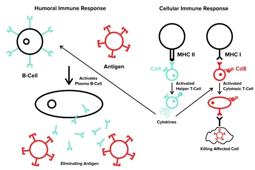
Figure 1 Humoral and cell-mediated immune responses.
-----
Part 5: Anatomy of the Immune System
We have learned of many different components of the immune system. In addition to anatomical barriers and white blood cells, there are other organs in the body often tested on the MCAT. Some of these components have been covered in previous sections of this chapter, but this section will cover additional macroscopic structures that help the immune system function. These include the bone marrow, the spleen, the thymus, and the lymphatic system.
a) Bone marrow
As you know, bone marrow is the site of hematopoiesis, or the production of blood cells. Since white blood cells are integral to the immune response, bone marrow is essential to keeping the immune system running properly. During hematopoiesis, stem cells divide into one of two subtypes: myeloid stem cells or lymphoid stem cells. Lymphoid stem cells are able to differentiate further into lymphocytes, while myeloid stem cells differentiate the remaining types of white blood cells, in addition to red blood cells (erythrocytes).
b) Spleen
The spleen can be thought of as a blood cell recycling center. It breaks down red blood cells and antibody-tagged bacteria and holds a reserve of blood.
The white pulp of the spleen is particularly important to the immune system. It has a large store of lymphocytes and is an activation site for B cells. It acts similarly to lymph nodes (more on that later) and plays an important role in coordinating the immune response through its high concentration of B and T cells.
c) Thymus
Once T cells are produced in the bone marrow, they are sent to the thymus to mature and differentiate. This is the site of positive and negative selection of T cells. The thymus is located in the center of the chest and is the namesake of T cells. (B cells were originally researched in birds and were found to mature in an organ called the bursa of Fabricius. Humans have no such organ, but the “B cell” name has stuck.)
The thymus plays little additional role in the immune response.
d) Lymphatic system
The lymphatic system plays an important role in both the circulatory and immune systems. In the circulatory system, the lymphatic system helps maintain fluid pressure throughout the body by draining the nutrient-rich interstitial fluid surrounding body tissue. The lymphatic system operates through its own network of lymph vessels that converge into lymph ducts and drain into the circulatory system at the subclavian vein.
Due to the presence of lymph vessels throughout the body and the lack of active pumping, the lymphatic system plays a large role in the passive equalization of fluid distribution. The lymphatic system also plays a large role in the absorption and transportation of proteins and lipid nutrients. (For more information on this, be sure to refer to our guide on amino acid and lipid metabolism.)
Throughout the body, there are hundreds of lymph nodes that serve as filters of lymph and are reservoirs of memory B and T cells. The lymphatic system includes the spleen and the thymus, as they are connected to the lymphatic circulation.
-----
Part 6: Antigens and Antibodies
Recall that B cells are responsible for producing antibodies. In this part, we will further discuss the structure and function of antibodies.
Antibodies will indicate to the body when an immune response is necessary. Thus, antibodies must be able to:
1) recognize foreign particles known as antigens
2) be recognizable by other components of the immune system
To understand how antibodies are able to execute these two simple functions, we must understand its structure.
a) Antibody structure
Human antibodies (also known as immunoglobulins) have a Y-shaped configuration made of two heavy chains and two light chains. The subunits are joined together by disulfide bonds, and within each antibody there is a variable region (where the antigen binds) and a constant region.
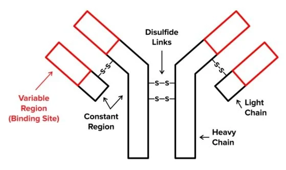
Figure 2 An antibody is composed of 2 heavy and 2 light chains. The heavy chains are linked by disulfide bonds. The light chains are connected to the heavy chains via disulfide bonds. The "tips" of the Y shape that an antibody posses are variable while the "bottom" of the "Y" shape is the constant region
There are 5 classes of immunoglobulins, differentiated by their heavy chains: IgA, IgD, IgE, IgG, and IgM. The constant regions are meant for recognition by other immune system cells.
Regarding the antigen, the site recognized by the antibody is known as the epitope. The terminal side of the antibodies—containing both the heavy and light chains—is composed of a hypervariable antigen-recognizing area (the result of random mutations during B cell maturation). The antigen-recognizing area of the antibody, known as the paratope, undergoes extensive random recombination to create a vast range of different antibodies in the body.
Table 1 Classes and functions of human immunoglobulins.
| Class of Immunoglobulin | Function | Location in the Body |
|---|---|---|
b) Antigen presentation
T cells recognize antigens through the help of major histocompatibility complexes (MHC’s). This section will go into greater detail about these proteins and their role in antigen presentation. There are two classes of MHCs:
MHC class I is a protein expressed on the surfaces of all nucleated cells in the human body. Its role is to take fragments of proteins within the cell and present them on the surface of the cell. When the cell is healthy, the protein being displayed will also be healthy and thus will elicit no response from the immune system. On the other hand, virally infected cells will present a viral protein. This viral protein will be recognized as an antigen and will elicit an immune response to kill those cells.
MHC class II is found in a smaller range of white blood cells that includes macrophages, dendritic cells, and B cells. These cells use machinery to destroy foreign objects in the body, displaying pieces of these foreign particles on MHC II.
Note that MHC I presents proteins that were internal to the cell, and MHC II presents proteins that were external to the cell.
Although all nucleated cells can present antigens through MHC I, the term antigen presentation is typically reserved for the action of cells with MHC class II. Antigen presentation by MHC II is a crucial part of the immune system because a foreign particle that is not presented will not elicit an immune response by T cells.
In the next section, we will discuss the recognition of self vs. non-self material within the body. Antigen presentation is also a key concept for various lab techniques (such as ELISA and Western blotting). These lab techniques use antigen presentation to visualize a protein upon applying compatible antibodies.
For further information on the role of major histocompatibility complexes in immune response, refer to the “T-cells and cytotoxic immunity” section above.
c) Recognition of self and non-self
While the immune system functions to eliminate foreign particles, it is just as important to make sure that immune cells only act on cells that are not supposed to be within the body: such as particles that were not part of the “self” or “self” cells that have become corrupted.
Recall that all B and T cells undergo a selection process checking that they do not bind and react to proteins of the self. Billions of immune cells survive this process, but many others do not. The cells that survive then proliferate when they bind to antigen and create “clones” of the reactive B and T cells—a process known as clonal selection. In hypersensitivity disorders, including autoimmune disorders and allergies, the negative selection process has failed to eliminate all T- and B- cells that react to the “self.”
Autoimmune disorders are exactly what they sound like: disorders in which the body thinks self cells are non-self and launches an immune response against itself. This can be incredibly dangerous. Examples of such disorders include lupus, rheumatoid arthritis, and multiple sclerosis (MS). You don’t need to know the specifics of these diseases, but it is important to understand how autoimmune disorders relate to the general immune system.
Note that the amount of response generated by the immune system is in balance with the well-being of the entire body.
If the immune system generates a critically low amount of response, the body is susceptible to infection.
If the immune system generates a critically high amount of response, such as in response to the self, the body is susceptible to autoimmunity.
The “right” amount of sensitivity is neither too high nor too little. It provides just the right amount of defense against foreign pathogens while refraining from attacking the cells of the self.
Allergies are another form of hypersensitive reaction by the immune system. They are typically not as severe as an autoimmune disorder but can have severe consequences, nonetheless. An allergic response occurs when there is an immune response mounted against a foreign particle that is normally regarded as harmless. These responses occur against a variety of items, including pollen, pet hair, and many different food types. The severity of allergic responses can significantly vary as well.
-----
Part 7: High-yield terms
Innate immune system: responsible for general disease protection and has no recognition of specific particles or diseases
Lysozyme: protein often found in saliva that aids in the breakdown of the bacterial cell wall
Leukocytes: white blood cells, the primary cell type of the overall immune system
Neutrophils: most abundant type of white blood cell; operates by phagocytosing bacteria; first responder to an infection
Lymphocytes: second most common type of white blood cell; 3 main types spanning both the adaptive and innate immune systems
Natural killer cells: type of lymphocyte that responds to virus/tumor infected cells to kill them and prevent the spread of disease
Monocytes: type of white blood cell that traverses the body tissues and can differentiate into two different types of cells
Dendritic cells: present antigens to T cells; type of differentiated monocyte
Macrophages: large cells that phagocytose many pathogens and particles; type of differentiated monocyte
Eosinophils: type of white blood cell that target parasitic infections
Basophils: type of white blood cell that is involved in the allergic response by releasing histamine and heparin
Mast cells: similar to basophils, except they are mostly found in mucous membranes and connective tissues
Phagocyte: a type of cell that engages in phagocytosis, which is the engulfment and destruction of one cell by another
Complement system: a signaling cascade of many proteins to tag pathogens to recruit phagocytes to destroy them
Opsonization: the term for the process of proteins tagging pathogens in the complement system; results in phagocytosis of tagged pathogens
Cytokines: type of signaling protein that aids in the coordination of the immune response by recruiting various cells
Inflammation: a response to an injury or pathogens that increases blood flow to an affected area to increase the number of immune cells able to responds quickly
Adaptive immune system: a subpart of the immune system that is able to change and adapt to the environment and learn to recognize and eliminate specific pathogens
Naïve B cells: type of B cells that have yet to see any antigen; typically, those that have just been produced in the bone marrow
Clonal selection: process by which B and T cells proliferate when they encounter a specific antigen, allowing for only the immune cells that are necessary to proliferate
Plasma cells: short-lived proliferation of naïve B cells that secrete many antibodies in response to an infection
Memory B cells: type of B cell proliferation that lies dormant until the same antigen is encountered once again, upon which the memory B cell proliferates into more plasma cells
Primary immune response: when a naïve B cell first encounters an antigen, the process of recruiting helper cells and secreting antibodies is a longer process
Secondary immune response: when subsequently exposed to an antigen, the immune response is much quicker due to the presence of memory B cells
Humoral immunity: property of the adaptive immune system that can be retained and transferred without the presence of cells; associated with B cells and production of antibodies
Cell-mediated immunity: property of the adaptive immune system that uses cells to execute the immune response; associated with T cells
Positive selection: elimination of T cells that do not appropriately respond to antigens and thus leave the body vulnerable to infection
Negative selection: elimination of T cells that respond too harshly, sometimes against healthy human cells (the “self”), thus causing an autoimmune disorder
Helper T cells: CD4+ cells, bind to MHC class II; recruit and activate B cells, killer T cells, and other cells of the immune system by releasing cytokines
Killer T cells: CD8+ cells, cytotoxic T cells, bind to MHC class I; kill an infected cell that it encounters presenting an infected protein
Major histocompatibility complex: protein on the surface of human cells that present various antigens; MHC class I is present in all cells and only elicits a response when the presented protein is not healthy, MHC class II is present in specific immune cells that present antigens after phagocytosing pathogens in the body
Antigen-presenting cells: type of immune cell that has MHC II; include macrophages, dendritic cells, and B cells
Hematopoiesis: production of blood cells in the bone marrow
Erythrocyte: red blood cell, which carries oxygen through the blood
Antigen: foreign particles that are recognized by antibodies
White pulp: lymphoid tissue of the spleen, holding reserved B and T cells
Immunoglobulins: human antibodies that have a Y shaped configuration; there 5 classes in the human body
Epitope: region on the antigen that is recognized by an antibody
Paratope: area of the antibody that recognizes the antigen; undergoes extensive random recombination in order to create a wide variety of antibodies that recognize many antigens
Autoimmune disorder: disorder where immune cells start an immune response against healthy human cells; examples include lupus, multiple sclerosis, and rheumatoid arthritis
-----
Part 8: Passage-based questions and answers
An experiment was conducted to analyze the effect of certain cytokines on the response of immunoglobulins M and G. Researchers chose to use the cytokine interleukin-6 for the experiment. IgM is responsible for the initial response to an antigen, while IgG is the most abundant antibody. Both of these antibodies can be found in the blood, while IgM is also located on the surface of B cells.
Experiment 1
Researchers used mice unable to produce IL-6 and control mice who were able to analyze the presence of IgM and IgG in their blood. A harmless protein was added to assess the immune response. The presence of these two antibodies 7 days after the injection is shown in the figure below.
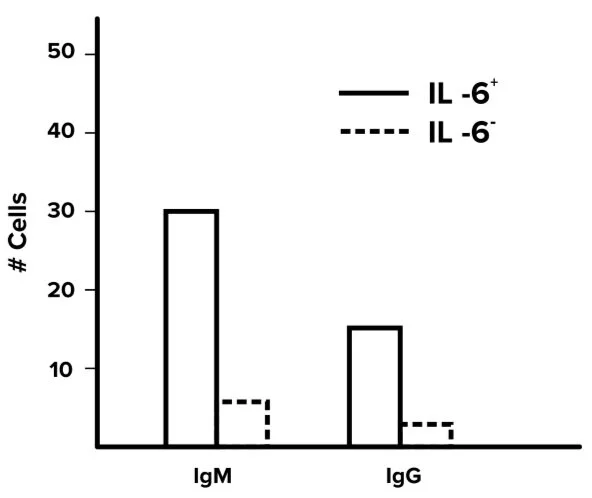
Figure 1 IgM and IgG quantities in treated mice.
Experiment 2
In an additional experiment, researchers visualized primary and secondary responses to an antigen using antibody titers over time for both IgM and IgG. IgM, a pentamer, has difficulty crossing blood vessels, but IgG, a monomer, is able to do so with ease. The results of this experiment are shown in the figure below.
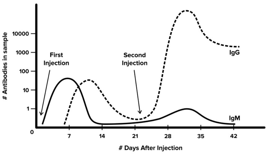
Figure 2 Experimentally determined levels of IgG and IgM in response to an antigen.
This passage was created for practice purposes only.
Question 1: Based on Figure 1 and Figure 2, which one of the following statements is most likely to be true?
A) IgM is insignificant in the immune response
B) IL-6 has no effect on the release of IgG
C) IgM responds earlier than IgG during an immune response
D) IgM plays the most significant role in the secondary immune response
Question 2: IgM and IgG are composed of what class of molecules?
A) Fatty acids
B) Amino acids
C) Carbohydrates
D) Leukocytes
Question 3: Which of the following is the most likely explanation of the results in Figure 2?
A) IgM moves throughout the body faster and responds more quickly than IgG
B) IgM serves to identify a new antigen and produce plasma cells
C) IgG recruits other cells of the immune system while IgM does not
D) The antigen that was used in the experiment is only identifiable by IgG
Question 4: Which of the following cell types are most likely to respond to IL-6?
A) Helper T cells and plasma B cells
B) Helper T cells and memory B cells
C) Cytotoxic T cells and plasma B cells
D) Cytotoxic T cells and memory B cells
Question 5: A “booster shot” for a vaccine would most likely have which effect?
A) Increase the formation of IgG
B) Inducing production of IgM, then IgG
C) Inducing production of IgG and IgM in similar quantities
D) A similar effect as an initial vaccine
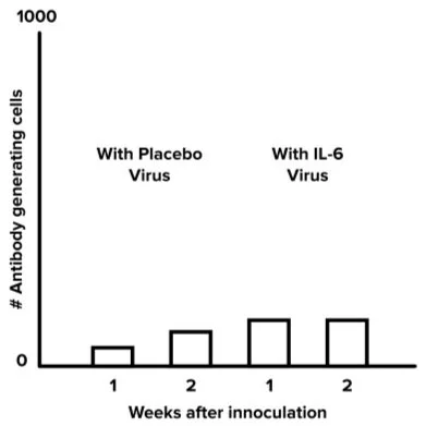
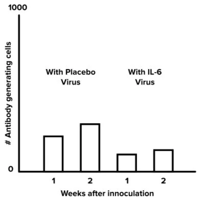
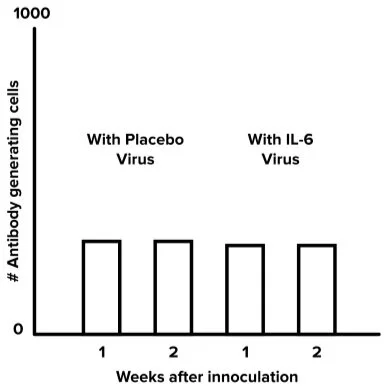
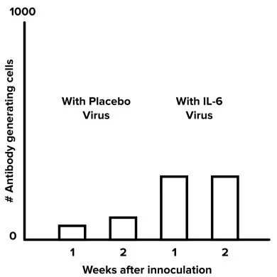
Question 6: If mice deficient in IL-6 were immunized with a synthetic virus containing IL-6, which of the following graphs would be most likely for the presence of antibody-secreting cells?
A)
B)
C)
D)
Answer key for passage-based questions
Answer choice C is correct. This question can be answered using the process of elimination. Figure 2 clearly shows IgM is the first one to spike after exposure to the antigen (choice A is incorrect). Even though IgG is lower than IgM, it can be seen in figure 2 that IgG just responds after IgM (choice B is incorrect). The secondary immune response is after the second injection of the antigen. Clearly, IgG has a higher peak than IgM (choice D is incorrect). There is a peak in IgM concentration, followed by a peak in IgG concentration, in the primary immune response.
Answer choice B is correct. Immunoglobulins are proteins, and proteins are made up of amino acids. Fatty acids make up lipids (choice A is incorrect). Carbohydrates include glucose and fructose and do not make up immunoglobulins (choice C is incorrect). Leukocytes refer to white blood cells (choice D is incorrect).
Answer choice A is correct. Figure 2 shows the levels of IgG and IgM over time. That IgM is only concerned with identifying a new antigen and creating plasma cells is untrue and does not explain why IgG has a higher peak (choice B is incorrect). If IgG were to recruit other cells, it would have a lower concentration and not display exponential growth in the secondary immune response (choice C is incorrect). If the antigen were only identifiable by IgG, there would be no response from IgM (choice D is incorrect).
Answer choice C is correct. IL-6 is a type of cytokine. Cytokines signal plasma cells to secrete more antibodies, as well as killer T cells to kill infected cells (choice C is correct).
Answer choice A is correct. A booster shot for a vaccine is equivalent to the antigen being introduced a second time (data in figure 2). Since it is the secondary immune response, we are concerned with the second half of the graph (choices B and C are incorrect). It is clear the first and second entrance of the antigen did not have the same effect (choice D is incorrect).
Answer choice D is correct. This question asks about a new experiment and what are the expected results. From figure 1, we see that the mice deficient in IL-6 do not produce either of the immunoglobulins significantly (choices B and C are incorrect). We also see that when IL-6 is present, the antibodies are produced in a significant quantity (choice A is incorrect).
-----
Part 9: Standalone practice questions and answers
Question 1: Which part of an antibody binds to antigens?
A) The constant region, consisting of only the heavy chain
B) The constant region, consisting of both the heavy and light chain
C) The variable region, consisting of both the heavy and light chain
D) The variable region, consisting of just the heavy chain
Question 2: A disease that impedes the functioning of MHC class II would have the least effect on which type of cell?
A) CD4+ cell
B) Macrophage
C) Dendritic cell
D) CD8+ cell
Question 3: Which of the following is not directly involved in the innate immune response?
A) Skin
B) Natural killer cells
C) Respiratory system
D) Thymus
Question 4: Which part of the immune system is most responsible for producing antibodies in response to an infection?
A) T cells in humoral immunity
B) B cells in humoral immunity
C) T cells in cell-mediated immunity
D) B cells in cell-mediated immunity
Question 5: What process prevents T cell-mediated autoimmune disorders?
A) Eliminating hyperactive immune cells through positive selection
B) Eliminating hyperactive immune cells through negative selection
C) Eliminating hypoactive immune cells through positive selection
D) Eliminating hypoactive immune cells through negative selection
Question 6: Which of the following processes facilitates the high variability and diversity in antibody structure?
A) Recombination of DNA
B) DNA methylation
C) Alternative splicing
D) Post-translational modification
Question 7: Which of the following is false regarding antibodies?
I. Antibodies tag antigens for destruction by killer T cells
II. Unique antibodies can be produced for every antigen
III. Antibodies can be passed from a mother to an infant
A) I, II, and III
B) III only
C) I and II only
D) II and III only
Answer key for standalone practice questions
Answer choice C is correct. Antibodies have both a heavy and a light chain, as well as a variable and a constant region. Antibodies bind to antigens in the variable region, which allows them to bind various antigens (choices A and B are incorrect). The paratope of the antibody is composed of both the heavy and light chains (choice D is incorrect).
Answer choice D is correct. MHC II is a protein only present on specialized antigen-presenting cells, which includes most white blood cells as well as helper T cells (choices A, B, and C are incorrect). This leaves CD8+ T cells, which are cytotoxic T cells that would be unaffected by a disease impairing MHC class II.
Answer choice D is correct. The thymus is primarily a storage for T cells, and thus plays a large role in the adaptive immune system. Skin tissue, natural killer cells, and dendritic cells are all nonspecific cells that aid in the innate immune response.
Answer choice B is correct. Antibodies are produced by plasma cells, which are a type of B cell (choices A and C are incorrect). While plasma cells are cells that secrete antibodies, B cells are part of the humoral immune response (choice D is incorrect).
Answer choice B is correct. Autoimmune disorders are caused by immune cells that attack self-cells, meaning they are hyperactive (choices C and D are incorrect). The process of eliminating these hyperactive cells is known as negative selection, while positive selection kills immune cells that do not elicit an immune response (choice A is incorrect).
Answer choice A is correct. DNA recombination is the process through which variable regions on antibodies are produced. DNA methylation is involved in epigenetics and gene silencing (choice B is incorrect). Alternative splicing refers to mRNA processing (choice C is incorrect). Post-translational modifications are on proteins after they are created and not how genetic variation is made (choice D is incorrect).
Answer choice C is correct. While antibodies tag antigens for destruction, killer T cells only kill human cells that present infected proteins. Option I is false (choices B and D are incorrect). While there is significant variation among antibodies, there is not an antibody for every single antigen, so option II is false as well. Antibodies can be passed from a mother to an infant through breast milk; option III is true (choice A is incorrect).